Canine Osteoarthritis Educational Series Part 1: What Causes Osteoarthritis?
Canine osteoarthritis (OA) is one of the most common conditions encountered in small animal practice, with prevalence of approximately 20% of dogs over one year of age, 40% of dogs over 4 years of age, and 80% of dogs over 8 years of age.1–4 Among certain at-risk breeds, prevalence exceeds 60%.4 For decades, OA was framed as a degenerative, mechanical wear-and-tear disease—an inevitable consequence of aging or use. That framing is outdated. Contemporary research portrays OA as a complex, whole-joint disease in which mechanical, inflammatory, metabolic, and immunologic processes converge to drive progressive structural failure of the osteochondral unit.5–8 Understanding this revised model matters clinically, because it reshapes how we counsel caregivers about prevention, when we intervene, and what we should expect from disease-modifying strategies.
Why “Wear and Tear” Doesn’t Tell the Whole Story
The wear-and-tear model assumes that cartilage simply erodes with use, much like brake pads on a car. If that were true, OA would be uniform across joints exposed to comparable loads, and protected joints would remain disease-free for life. Neither is the case. OA is now understood to be the clinical and pathological endpoint of a range of joint disorders driven by a dynamic imbalance between cartilage growth and breakdown, low-grade synovial inflammation propagated by immune dysregulation, subchondral bone remodeling, and altered mechanosensitive cellular signaling.
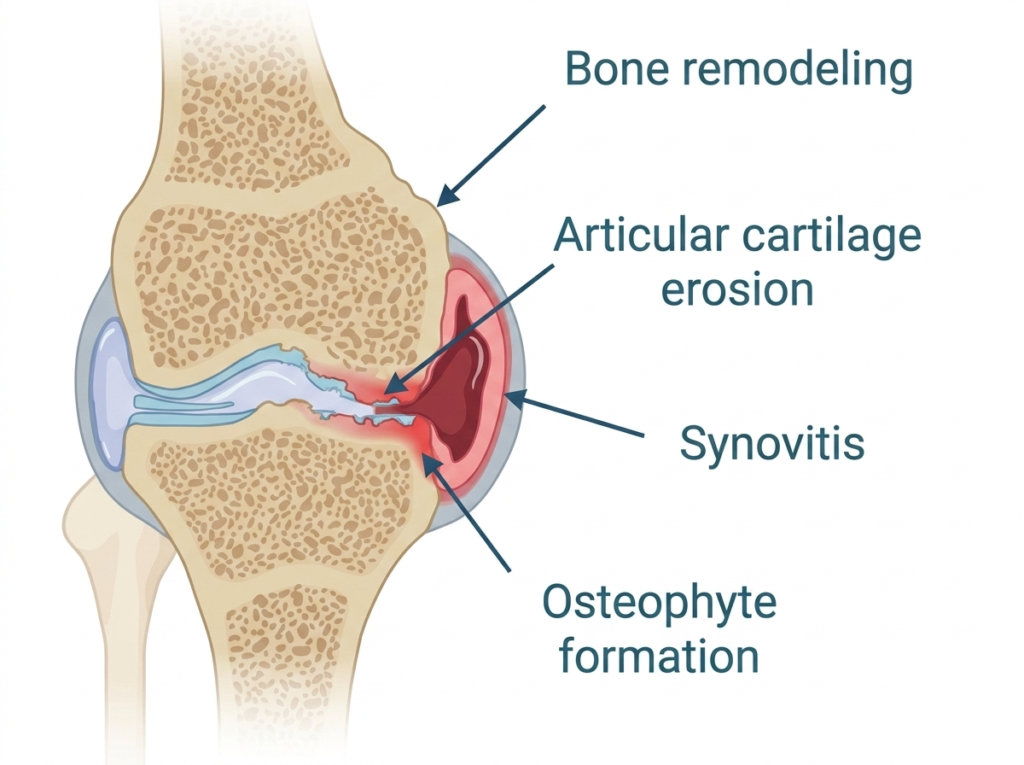
Articular cartilage (a type of hyaline cartilage) is composed primarily of water (70–80%), type II collagen, and proteoglycans, with chondrocytes representing only 1–2% of tissue volume.9 This avascular, aneural tissue depends almost entirely on synovial fluid diffusion for nutrition and has minimal intrinsic regenerative capacity.9 When even minor cartilage injury occurs, the repair response typically yields fibrocartilage rich in type I collagen rather than true hyaline cartilage, resulting in a tissue with inferior compressive strength, elasticity, and wear resistance.9 This biological reality means that joints have little tolerance for the cycle of microdamage and aborted repair that defines OA.
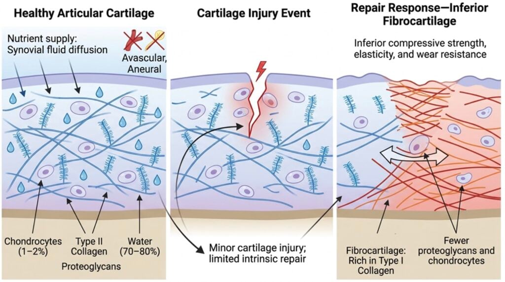
Importantly, the trigger for OA is rarely cartilage damage in isolation. In larger animal models of OA induced by meniscal injury, the earliest detectable changes occur in the subarticular spongiosa-trabecular bone. Degradation of structural architecture, loss of bone volume, and reduced mineralization precede measurable changes in the overlying subchondral bone plate or articular cartilage.10 This finding overturns the older view that OA “starts in the cartilage.” It begins in the entire osteochondral unit, with bone, cartilage, synovium, and joint capsule all participating from the outset.
The Mechanical-Inflammatory Loop: How OA Actually Develops
OA can be productively understood as the outcome of either abnormal forces acting on a normal joint, or normal forces acting on an abnormal joint.6 In either scenario, the inciting event is most often joint instability, incongruity, or an injury/trauma that creates a focal cartilage injury. What follows is a self-amplifying loop of mechanical and inflammatory signaling.
Mechanosensitive Signaling and the Chondrocyte Response
Chondrocytes are exquisitely mechanosensitive. Abnormal mechanical loading whether from joint laxity, malalignment, instability, or an osteochondral fragment, triggers altered intracellular signaling, increased cell death, and upregulation of catabolic enzymes.6,8 Once these mechanosensitive pathways are activated, they amplify the inflammatory cascade and the disease becomes self-perpetuating.6 The goals of disease modification are to reduce the inflammatory cascade and reestablish intra-articular cellular regulation that supports tissue repair before the inflammatory cascade becomes established.
The Synovial Inflammatory Cascade
Cartilage breakdown products released into the synovial fluid act as damage-associated molecular patterns (DAMPs), scavenged by type A synovial macrophages, which initiates synovitis.8 The synovial membrane develops villous hypertrophy and proliferation. Activated synoviocytes and chondrocytes secrete pro-inflammatory cytokines, most importantly interleukin-1β (IL-1β) and tumor necrosis factor-α (TNF-α), which in turn upregulate matrix metalloproteinases (MMPs), nitric oxide, and reactive oxygen species.6,9,11 MMP-13 in particular is implicated in the enzymatic destruction of type II collagen11, while ADAMTS family enzymes degrade aggrecan.
These cytokines promote macrophage polarization toward a tissue-destructive M1 phenotype and upregulate endothelial adhesion molecules, creating a chemotactic environment that recruits circulating immune cells into the joint.12 As the adaptive immune response becomes engaged, autoreactive T cells and autoantibody-producing B cells infiltrate the synovium, further amplifying and sustaining the inflammatory milieu.11,13
This process has been particularly well characterized in the canine stifle affected by cranial cruciate ligament disease. Histological studies of synovial tissue demonstrate infiltration by macrophages, T lymphocytes, B lymphocytes, and IgG-producing plasma cells in a pattern that closely resembles rheumatoid arthritis.14 As the cruciate ligament undergoes degeneration, released type I collagen acts as an intra-articular antigen, stimulating local anti-collagen antibody production and immune complex formation. This autoimmune-like response perpetuates chronic synovitis and joint inflammation and has been detected before clinical ligament rupture occurs.14
Prostaglandin E2 (PGE2) production can increase up to 50-fold in inflamed joints, further driving inflammation, sensitizing nociceptors, and stimulating MMP production.15 Toll-like receptors (TLR-2 and TLR-4) are upregulated on chondrocytes in OA joints, activating additional catabolic pathways.11 The synovial fluid simultaneously loses hyaluronic acid concentration and viscoelasticity, compromising its protective and lubricating functions.8
Each step reinforces the others, and the joint cannot easily return to baseline once the loop is established.
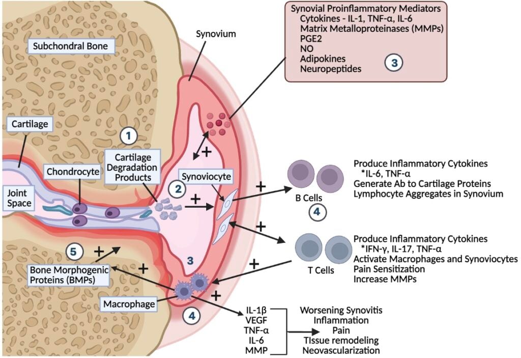
Adipose tissue is not metabolically inert. Adipokines (cytokines produced predominantly by adipose tissue but also locally within joints) play a pivotal and increasingly recognized role in OA pathogenesis.8,16 The major adipokines implicated in joint disease include leptin, adiponectin and several others. These molecules help regulate cartilage growth and breakdown, cartilage cell survival and death, subchondral bone remodeling, and inflammation.8
Leptin has emerged as a particularly important player.17 In dogs, serum and synovial leptin concentrations correlate with joint pain and dysfunction, and studies demonstrate that the severity of OA correlates with plasma and synovial leptin concentrations.8 Mechanistically, leptin promotes cartilage breakdown, increases chondrocyte cell death, and stimulates MMP production.8 Because adipose tissue is the primary source of circulating leptin, obesity directly increases the inflammatory burden on every joint in the body, whether or not those joints are mechanically overloaded. Adiponectin, visfatin, resistin, and chemerin contribute additional layers of complexity, with effects ranging from pro-inflammatory to immunomodulatory depending on context.8
Subchondral Bone: An Underappreciated Driver
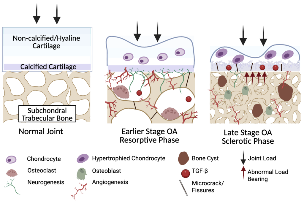
The subchondral bone plate and underlying trabecular spongiosa are active participants in OA, not passive bystanders. Early in disease, primary osteoporotic-like changes occur: increased subchondral bone plate porosity, decreased bone mineral density, loss of trabecular volume, and reduced complexity of the trabecular network.10 These changes are thought to result from microdamage to trabeculae under altered loading conditions.10 Soon thereafter, the previously normal subchondral bone plate becomes thicker; its porosity first increases, then decreases as sclerosis develops.10 Osteophyte formation at joint margins follows endochondral ossification pathways and represents an adaptive attempt to increase joint contact area and stability—at the cost of joint range of motion.8
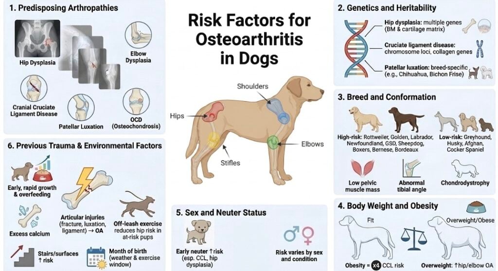
Risk Factors for Canine OA
Risk factors for canine OA and its predisposing arthropathies can be classified into six principal categories: genetics, breed, conformation, body weight, age, and sex/neuter status.1 These factors interact with environmental and lifestyle exposures to determine whether, when, and how severely a given dog will develop OA. Some of these risks are modifiable while others are not, but even the unmodifiable ones can be used to identify at-risk individuals for earlier surveillance and intervention.
1. Predisposing Arthropathies: The Most Common Pathway
Secondary OA arising from an identifiable underlying joint disease is far more common in dogs than truly idiopathic primary OA.18 The predisposing arthropathies that account for the majority of canine OA cases include:
- Hip dysplasia: Multifactorial polygenic disease characterized by joint laxity, subluxation, and abnormal joint development. Developmental contributors to OA will be discussed in greater detail in Part 2 of this series: “Developmental Contributors of OA.”
- Elbow dysplasia: An umbrella term encompassing medial coronoid disease, radioulnar incongruity, ununited anconeal process, and osteochondritis dissecans of the medial humeral condyle. Medial coronoid disease is now the most common cause of forelimb lameness in young large-breed dogs and is characterized by subchondral osteonecrosis, cartilage lesions, and vasculitis. This topic will be discussed in more detail in Part 2 of this series.
- Cranial cruciate ligament disease: Unlike acute traumatic anterior cruciate ligament rupture in human athletes, canine cruciate disease is a degenerative process with progressive ligament insufficiency, low-grade synovitis, and early OA onset. Symptoms may remain subclinical for extended periods before acute decompensation. This topic will be discussed in more detail in “Canine OA Educational Series 3: Stifle Disease.”
- Patellar luxation: Most often medial luxation in small breeds (Pomeranian, Chihuahua, Yorkshire Terrier, French Bulldog), but lateral and large-breed luxations also occur. Severity is graded I–IV; even grade I and II luxations can lead to OA over time. This topic will be discussed in more detail in Part 3 of this series.
- Osteochondrosis / Osteochondritis dissecans (OCD): Disturbance of endochondral ossification with vascular failure in the secondary ossification center. Most commonly affects the shoulder (humeral head), elbow, stifle, and tarsus in growing large-breed dogs. This topic will be discussed in more detail in Part 2 of this series.
Each of these conditions produces joint instability or incongruity, which initiates the mechanical-inflammatory loop described above. The earlier the underlying arthropathy is identified and addressed, the better the long-term joint outcome.18
2. Genetics and Heritability
Genetic predisposition is the most frequently reported risk factor in the canine OA literature.1 Hip dysplasia heritability estimates cluster around 0.28–0.57, with multiple candidate genes identified including those involved in basement membrane and cartilage matrix proteins.1 For cranial cruciate ligament disease, genome-wide association studies have identified loci on chromosomes 1, 3, 5, 13, 24, and 33, with collagen genes prominently implicated1. Patellar luxation heritability has been estimated at 0.21–0.44 in Chihuahuas, Bichon Frise, and Pomeranians1. Despite decades of breeding interventions, prevalence in affected breeds has remained relatively static, indicating that genetic complexity and strong environmental modifiers continue to challenge simple phenotypic selection strategies.
3. Breed and Conformation
Breed risk reflects the combined contributions of genetic background, body size, and conformational standards. Breeds with significantly increased OA risk include Rottweilers, Golden and Labrador Retrievers, Newfoundlands, German Shepherds, Old English Sheepdogs, Dogue de Bordeaux, Boxers, and Bernese Mountain Dogs1. Smaller breeds and certain working lines—notably greyhounds, Siberian Huskies, Afghan Hounds, and Cocker Spaniels—show lower OA risk.1
Conformational traits with documented OA associations include low pelvic muscle mass (hip dysplasia and OA risk), tibial tuberosity width and proximal tibial angle (cruciate ligament disease risk), and chondrodystrophic limb conformation (elbow incongruity).1,8 Selection pressures for extreme conformational traits have inadvertently co-selected for orthopedic vulnerability in several modern breeds.
4. Body Weight and Obesity
Excess body weight is among the most powerful modifiable risk factors for canine OA. In every published study examining body weight as a risk factor, increased weight was associated with increased OA risk.1 Obesity nearly quadruples the risk of cranial cruciate ligament disease (odds ratio ~3.8), and overweight body condition is consistently associated with elbow and hip OA.1
The seminal Labrador Retriever lifetime restricted-feeding study demonstrated that dogs maintained at a lean body condition from puppyhood lived on average 1.8 years (15%) longer than their free-fed littermates.19 At the end of the study, 68% of the heavier dogs had hip or shoulder OA compared to only 10% of the lean dogs, and 77% of heavier dogs had OA in two or more joints compared to 10% of lean dogs.19 The mechanisms are dual: increased mechanical load on joints, and increased systemic inflammatory tone mediated by adipokines.8 This dual mechanism explains why even modest weight reduction can produce disproportionate clinical improvement.
4. Sex and Neuter Status
Neutered dogs are consistently reported to have higher OA risk than intact dogs across multiple studies and joint diseases.1 The mechanisms are not fully resolved and likely involve several factors: post-neutering weight gain, altered gonadal hormone exposure during skeletal development, and timing of growth plate closure.1,20 Early neutering (before skeletal maturity) has been particularly implicated in increasing the risk of cruciate ligament disease and hip dysplasia in some breeds, notably Golden Retrievers.1 Sex effects are less consistent across studies, with some reporting increased risk in females and others in males depending on the specific condition. These findings underscore that neutering decisions should be individualized considering breed, body size, sex, and orthopedic risk profile rather than applied by a single default age.
5. Previous Trauma and Joint Injury
Any joint injury, whether a single acute event or cumulative microtraumas, can initiate OA. This is particularly relevant in working and sporting dogs that begin athletic training before skeletal maturity.20 Articular fractures, joint luxation, ligament injuries, and osteochondral fragmentation all create the joint instability or surface incongruity that triggers the mechanical-inflammatory cascade. Even successfully managed surgical cases almost invariably progress to some degree of OA over time,20 which is why post-injury joint surveillance and long-term multimodal management are so important. The pathophysiology of OA in working dogs and canine athletes will be discussed in greater detail in “Canine OA Educational Series 4: Osteoarthritis in the Canine Athlete.”
6. Early Life and Environmental Factors
Several lifestyle and developmental factors modify OA risk. Rapid growth and overfeeding during puppyhood, particularly in large and giant breeds, accelerate skeletal development in ways that predispose to hip dysplasia, elbow dysplasia, and developmental orthopedic disease.20 Excess calcium intake (above approximately 4.5 g per 1000 kcal) is similarly implicated.20 Counterintuitively, off-leash exercise on appropriate terrain in young, at-risk puppies has been associated with lower hip dysplasia clinical signs in some studies, whereas exposure to stairs and certain surfaces increased risk.1 Month of birth has been associated with OA risk in some populations, likely reflecting exposure to early-life exercise opportunities based on weather conditions.1 These findings collectively support a model in which appropriate early-life physical activity supports joint development, while either insufficient or excessive activity and overnutrition predispose to disease.
How OA Pathophysiology Changes with Age
OA is sometimes treated as a single disease state, but its biology shifts substantially across the canine lifespan. Recognizing these stage-specific differences is critical for clinical management.
OA in Growing Dogs: An Acute, Inflammatory Phenotype
In puppies, OA most often arises as a consequence of developmental joint disease including hip dysplasia, elbow dysplasia, OCD, or patellar luxation.20 The dominant pathological features are acute inflammation and acute pain rather than chronic structural changes. Subchondral bone inflammation, joint capsule strain from instability, and cartilage damage at point-loading sites dominate the early picture.20 Clinically, puppies with OA tend to show transient lameness, subtle behavioral changes (reduced playfulness, mild introversion), and pain on joint manipulation.20 Importantly, loss of muscle mass, contracture, and significantly restricted joint motion (features of chronic OA) are uncommon at this stage.
This phenotype matters because acute inflammatory pain responds well to multimodal management and may be at least partially reversible if the underlying instability is addressed.20 Once physical changes to the joint structure and chronic central and peripheral pain sensitization develop, the neurophysiologic signature becomes more complex and treatment requirements expand substantially.
OA in the Adult Dog: Established Disease and Sensitization
By adulthood, dogs with predisposing arthropathies typically have established radiographic OA characterized by osteophytes, subchondral sclerosis, joint capsule thickening, and synovial proliferation.10 This topic will be discussed in greater detail in “Canine OA Educational Series 6: Screening Radiographs by Life Stage.” The inflammatory tone of the joint persists at a lower grade, but the disease landscape is now dominated by structural changes—loss of joint range of motion, periarticular fibrosis, muscle mass loss from disuse, and altered gait mechanics that further compromise joint biomechanics.8 Pain becomes chronic, with associated peripheral and central sensitization that lower the threshold for nociception. The relationship between radiographic findings and pain presentation will be discussed in greater detail in Part 8 of this series. Adipokine-mediated systemic inflammation continues to drive disease progression, particularly in overweight individuals.
OA in the Senior Dog: Chondrocyte Senescence and Multi-Joint Disease
Aging exerts independent effects on cartilage biology that compound any preexisting OA. With age, cartilage cell numbers decline, the remaining cells show signs of aging and altered cartilage production, and they become more prone to cell death.9 The capacity for matrix repair diminishes when accumulated mechanical and inflammatory damage is greatest. Senior dogs commonly have multi-joint OA, often combined with comorbid musculoskeletal and neurologic conditions (intervertebral disc disease, lumbosacral stenosis, muscle loss), which complicates pain localization and management. The neurophysiologic signature of chronic pain in senior dogs is further altered by age-related changes in central pain processing. This topic will be discussed in greater detail in “Canine OA Educational Series 5: Osteoarthritis in the Geriatric Dog.”
Clinical Implications: Putting the Pathophysiology to Work
The reframing of OA from a passive wear-and-tear process to an active, multi-factorial, whole-joint inflammatory disease has direct clinical implications:
- OA in dogs is rarely idiopathic. In most cases, an identifiable predisposing arthropathy (dysplasia, ligament disease, luxation, OCD) is driving the disease. Identifying and addressing the underlying mechanical problem early, before the inflammatory cascade becomes self-sustaining, offers the best chance of disease modification.
- Body weight is not a cosmetic issue. Adipose tissue is an active inflammatory organ that drives OA through both mechanical and adipokine-mediated mechanisms. Lean body condition is one of the most powerful disease-modifying interventions available, with effect sizes comparable to or exceeding many pharmacologic options.
- Subchondral bone is an active participant in OA from the earliest stages. Therapeutic strategies aimed exclusively at cartilage miss a major component of the disease.
- Age changes the disease, not just its severity. Puppy OA, adult OA, and senior OA require different management priorities, and clinicians should communicate these stage-specific expectations clearly to owners.
Genetic and breed risk identifies at-risk dogs early. While the alleles themselves cannot yet be modified clinically, knowing a dog’s baseline risk profile permits proactive screening, growth and weight management, and targeted owner education.
Clinically, the signs most often recognized in animals with late-stage osteoarthritis are the result of a prolonged inflammatory process. Over time, this persistent inflammatory microenvironment drives both peripheral and central sensitization, culminating in a maladaptive chronic pain state that becomes the most debilitating feature of advanced disease.
While management of these pain pathways is critically important, the more important lesson is that intervention should not begin when chronic pain becomes apparent. Veterinarians and pet owners have valuable opportunities to act earlier in the disease course, when therapies aimed at modifying joint pathology may help prevent or delay the progression to irreversible damage and chronic pain.
OA is no longer the inevitable, untreatable end-of-life condition it was once thought to be. For veterinary clinicians, the modern understanding of OA pathophysiology offers something genuinely useful: identifiable mechanisms, modifiable risk factors, and meaningful windows for intervention across the lifespan of the dog.
Stay tuned for next month’s “Canine Educational Series 2: Developmental Contributors of Osteoarthritis.”
References
- Anderson KL, Zulch H, O’Neill DG, Meeson RL, Collins LM. Risk Factors for Canine Osteoarthritis and Its Predisposing Arthropathies: A Systematic Review. Front Vet Sci. 2020;7:220. doi:10.3389/fvets.2020.00220
- O’Neill DG, Church DB, McGreevy PD, Thomson PC, Brodbelt DC. Prevalence of Disorders Recorded in Dogs Attending Primary-Care Veterinary Practices in England. PLoS One. 2014;9(3):e90501. doi:10.1371/journal.pone.0090501
- Anderson KL, O’Neill DG, Brodbelt DC, et al. Prevalence, duration and risk factors for appendicular osteoarthritis in a UK dog population under primary veterinary care. Sci Rep. 2018;8(1):5641. doi:10.1038/s41598-018-23940-z
- Enomoto M, de Castro N, Hash J, et al. Prevalence of radiographic appendicular osteoarthritis and associated clinical signs in young dogs. Sci Rep. 2024;14(1):2827. doi:10.1038/s41598-024-52324-9
- Meeson RL, Todhunter RJ, Blunn G, Nuki G, Pitsillides AA. Spontaneous dog osteoarthritis — a One Medicine vision. Nat Rev Rheumatol. 2019;15(5):273-287. doi:10.1038/s41584-019-0202-1
- Tomé I, Alves-Pimenta S, Sargo R, et al. Mechanical osteoarthritis of the hip in a one medicine concept: a narrative review. BMC Vet Res. 2023;19(1):222. doi:10.1186/s12917-023-03777-z
- Marcellin-Little DJ, Hulse DA, Huntingford JL, et al. A proposed framework for practical multimodal management of osteoarthritis in growing dogs. Front Vet Sci. 2025;12:1565922. doi:10.3389/fvets.2025.1565922
- Theyse LFH, Mazur EM. Osteoarthritis, adipokines and the translational research potential in small animal patients. Front Vet Sci. 2024;11:1193702. doi:10.3389/fvets.2024.1193702
- Thoene M, Bejer-Olenska E, Wojtkiewicz J. The Current State of Osteoarthritis Treatment Options Using Stem Cells for Regenerative Therapy: A Review. Int J Mol Sci. 2023;24(10):8925. doi:10.3390/ijms24108925
- Oláh T, Cucchiarini M, Madry H. Subchondral bone remodeling patterns in larger animal models of meniscal injuries inducing knee osteoarthritis – a systematic review. Knee Surg Sports Traumatol Arthrosc. 2023;31(12):5346-5364. doi:10.1007/s00167-023-07579-6
- Haseeb A, Haqqi TM. Immunopathogenesis of osteoarthritis. Clin Immunol. 2013;146(3):185-196. doi:10.1016/j.clim.2012.12.011
- Mathiessen A, Conaghan PG. Synovitis in osteoarthritis: current understanding with therapeutic implications. Arthritis Res Ther. 2017;19(1):18. doi:10.1186/s13075-017-1229-9
- Sanchez-Lopez E, Coras R, Torres A, Lane NE, Guma M. Synovial inflammation in osteoarthritis progression. Nat Rev Rheumatol. 2022;18(5):258-275. doi:10.1038/s41584-022-00749-9
- Doom M, De Bruin T, De Rooster H, Van Bree H, Cox E. Immunopathological mechanisms in dogs with rupture of the cranial cruciate ligament. Vet Immunol Immunopathol. 2008;125(1-2):143-161. doi:10.1016/j.vetimm.2008.05.023
- Trumble TN, Billinghurst RC, McIlwraith CW. Correlation of prostaglandin E2 concentrations in synovial fluid with ground reaction forces and clinical variables for pain or inflammation in dogs with osteoarthritis induced by transection of the cranial cruciate ligament. Am J Vet Res. 2004;65(9):1269-1275. doi:10.2460/ajvr.2004.65.1269
- Kleine SA, Sanderson SL, George C, et al. Correlation of serum and synovial leptin concentrations with body condition scores in healthy and osteoarthritic dogs. Vet Surg. 2019;48(5):780-785. doi:10.1111/vsu.13244
- Carrión M, Frommer KW, Pérez-García S, Müller-Ladner U, Gomariz RP, Neumann E. The Adipokine Network in Rheumatic Joint Diseases. Int J Mol Sci. 2019;20(17):4091. doi:10.3390/ijms20174091
- Johnston SA. Osteoarthritis. Joint anatomy, physiology, and pathobiology. Vet Clin North Am Small Anim Pract. 1997;27(4):699-723. doi:10.1016/S0195-5616(97)50076-3
- Kealy RD, Lawler DF, Ballam JM, et al. Effects of diet restriction on life span and age-related changes in dogs. J Am Vet Med Assoc. 2002;220(9):1315-1320. doi:10.2460/javma.2002.220.1315
- Mosley C, Edwards T, Romano L, et al. Proposed Canadian Consensus Guidelines on Osteoarthritis Treatment Based on OA-COAST Stages 1–4. Front Vet Sci. 2022;9:830098. doi:10.3389/fvets.2022.830098

Dr. Marie Bartling is a veterinarian with more than 20 years of clinical experience specializing in integrative medicine, pain management, rehabilitation, and regenerative therapies.
Her approach combines conventional medicine with advanced therapies to manage complex medical conditions, musculoskeletal disease, and chronic pain. She is passionate about translating emerging science into practical, evidence-based strategies that improve patient health, mobility, and quality of life.

Vice President of Veterinary Affairs at Gallant
Dr. Rebecca Windsor, DVM, DACVIM, is a board-certified veterinary neurologist with over 20 years of clinical experience and a strong record of scientific publication. She joined Gallant in 2025 and serves as Vice President of Veterinary Affairs.
Dr. Windsor specializes in veterinary regenerative medicine, with a focus on advancing FDA-approved, off-the-shelf mesenchymal stromal cell (MSC) therapies for pets. She develops educational platforms that translate the science, safety, and clinical efficacy of stem cell therapy for veterinary professionals. Since 2019, she has served as a Clinician Scientist at Ethos Discovery, where she leads the neurology research portfolio.