The Lungs are the Point: Rethinking Pulmonary “Trapping” of Intravenous Mesenchymal Stromal Cells
If you have read about intravenous (IV) mesenchymal stromal cell (MSC) therapy, chances are you have encountered some version of this concern: “most of the cells just get trapped in the lungs and never reach the target tissue.” We now know that this statement isn’t true, or perhaps relevant. Yes, the lungs temporarily hold the majority of IV-administered MSCs. However, this pulmonary trapping is not a therapeutic failure — it turns out to be the mechanism through which the therapy works. A careful look at the scientific literature, beginning with the influential 2009 study that first framed pulmonary trapping as a “problem,” reveals a story of science catching up with itself: what was once characterized as an obstacle has been reinterpreted, step by step, as the engine of efficacy.
What Is the Pulmonary First-Pass Effect?
The pulmonary first-pass effect describes the initial localization of intravenously administered cells within the pulmonary microvasculature before they can enter systemic arterial circulation. For MSCs, this early retention reflects a combination of cell size, deformability, dose and infusion conditions, and interactions with the pulmonary endothelium. The terms pulmonary trapping, pulmonary retention, lung entrapment, and pulmonary first-pass effect are often used interchangeably, although “retention” is generally the most neutral description.
The Study That Started the Conversation
In 2009, an MSC biodistribution study in rats was published titled “Pulmonary Passage Is a Major Obstacle for Intravenous Stem Cell Delivery.”1 In this study, tracker-labeled MSCs and other cell types were infused into the jugular vein of anesthetized rats, and arterial blood was sampled continuously from the carotid artery to count how many cells crossed into the arterial system within 10 minutes. Infrared imaging of harvested organs showed bright lung signal in nearly every group, with no detectable signal in any other organ.
The conclusion – that pulmonary trapping represents a “major obstacle” to IV stem cell delivery – has been cited hundreds of times since and has shaped how clinicians and scientists think about the route of delivery for MSC therapy. However, careful reading of the study and an understanding of how the field has evolved in the nearly 20 years since publication reveals that this framing rests on a series of assumptions that simply do not hold in the clinical context where IV MSC therapy is used.
What the Study Could Not Determine
1. It Used Healthy Rats Without an Active Disease Target
The most important limitation of the study, one the authors themselves acknowledged, but that has largely been lost in the paper’s subsequent citation history: the rats were healthy. They were anesthetized with isoflurane, had no injury, no inflammation, and no pathological process generating chemotactic signals that would recruit circulating cells out of the pulmonary vasculature and into tissue.
In the inflammatory environment of a real patient, upregulation of pro-inflammatory cytokines (i.e., IFN-γ and TNF-α) and chemokines act as homing signals that modify where and how MSCs distribute. This signaling environment facilitates redistribution from the lungs over time and activates MSCs, “licensing” them to secrete anti-inflammatory molecules at far higher levels than quiescent cells would produce.
Testing pulmonary passage of stem cells in a healthy, anesthetized rat and concluding that cells “cannot reach the target organ” is equivalent to assessing whether firefighters can reach a fire by observing them at the fire station and noting that they are not moving—simply because they have never received a call to respond. The biological machinery that moves cells to sites of inflammation is simply not running in a homeostatic animal.
2. The Imaging Could Not Detect Small Numbers of Distributed Cells
The infrared imaging used in the study showed bright lung signal and apparently no signal in other organs, leading to the conclusion that cells could not be detected elsewhere. But macroscopic infrared imaging of whole harvested organs is a relatively insensitive detection method. Advancements in radiolabeling techniques have improved sensitivity to detect MSCs in the tissues.2
3. The Study Measured Immediate Passage, Not Longer-Term Biodistribution or Efficacy
The study measured the percentage of cells crossing into the carotid artery within 10 minutes of infusion.1 This is a valid measure of immediate pulmonary passage, but it does not capture later MSC biodistribution, cell clearance, immune interactions, or therapeutic outcomes that may unfold over the following 24 to 72 hours.
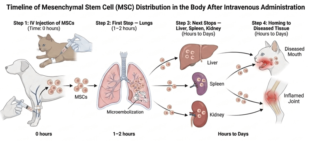
Multiple subsequent studies have tracked radiolabeled or fluorescently tagged MSCs over longer timeframes and documented a consistent, reproducible pattern: cells are predominantly lung-retained in the first 1–2 hours, then gradually redistribute to the liver, spleen, kidney, and injured target tissues over the course of 24–48 hours.2–4 One study of a canine myocardial infarction model documented focal cardiac homing visible within 24 hours of IV infusion that persisted for 7 days.5 The 10-minute arterial sampling in healthy rats cannot speak to any of this.
More importantly, the therapeutic question — whether enough cells reach the target tissue to produce benefit — may not even be the right question. As the field has come to understand, the mechanism of IV MSC therapy does not primarily depend on cells arriving at and engrafting in diseased tissue at all.
Does Pulmonary Trapping Mean IV MSCs Cannot Work?
No. Pulmonary retention demonstrates that many intact cells do not immediately pass through the lungs into systemic arterial circulation. It does not, by itself, establish that IV MSC therapy is ineffective. Depending on the cell product and disease model, therapeutic effects may be mediated through paracrine signaling, inflammatory licensing, immune-cell interactions, apoptosis and efferocytosis, extracellular vesicles, or the redistribution of a smaller population of MSCs.
Why Do IV MSCs Initially Accumulate in the Lungs?
Far from being a passive trap, the lung is an active site of MSC education, activation, and therapeutic signaling. Understanding what happens to MSCs during their pulmonary residence reframes the conversation about IV MSC delivery.
Cell Size and Receptor-Mediated Adhesion
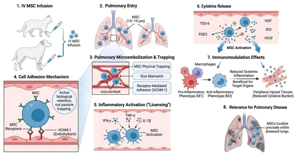
MSCs are large cells, typically 15–19 μm in diameter, which is larger than the diameter of pulmonary capillaries. This size mismatch means that cells form microemboli in the pulmonary microvasculature upon IV infusion — a physical reality that has been confirmed across species.6,7
But size is not the only factor. Receptor-mediated adhesion plays a critical role, and blocking surface receptors on MSCs can significantly increase the proportion of cells crossing into the arterial circulation. VCAM-1 on the pulmonary vascular endothelium appears to be the primary binding partner, facilitating active adhesion in addition to the passive size-based trapping. This means that pulmonary retention is a biologically regulated process, not merely a mechanical accident, and that the lung actively participates in retaining these cells.
MSCs in the Lungs Are Activated, Not Dormant
Once retained in the pulmonary vasculature, MSCs are not passive. Many studies have demonstrated that activated lung-trapped cells alter the systemic inflammatory environment to ultimately benefit the target organ. The pulmonary entrapment combined with inflammatory signals from the systemic circulation (i.e., IFN-γ, TNF-α, IL-1β) activates or “licenses” the cells to upregulate expression of anti-inflammatory mediators to levels far higher than unstimulated cells would produce.8,9 These effects reflect the broader immunomodulatory properties of mesenchymal stromal cells, including their interactions with the recipient’s innate and adaptive immune systems.
MSCs in the lung have been shown to upregulate expression of TSG-6 (tumor necrosis factor-stimulated gene 6), a potent anti-inflammatory glycoprotein that has been shown to improve cardiac function in mouse infarction models.10 Lung trapped cells also upregulate PGE2, IL-10, TGF-β, HGF, IDO, and VEGF, a potent collection of molecules that dampen systemic inflammation, shift macrophage polarization, and reduce the cytokine burden in injured tissues throughout the body.11 In conditions where the lungs themselves are diseased, pulmonary trapping delivers cells precisely where they are needed.
Where Do Cells Go After the Lungs?
Pulmonary retention is temporary. Beginning within hours of infusion, cells begin to clear from the lungs and redistribute to the liver, spleen, kidney, and diseased tissue(s).2,3,12,13 When injury-specific signals are present, redistribution is not random. The SDF-1/CXCR4 axis is the best-characterized homing mechanism: SDF-1 is upregulated at sites of tissue injury and circulating MSCs expressing CXCR4 migrate toward this gradient. This has been demonstrated experimentally in models of myocardial infarction, stroke, kidney injury, liver injury, and bone marrow irradiation.
The Most Surprising Discovery: Dead Cells Keep Working
Perhaps the most paradigm-shifting finding in modern MSC biology is that the death of infused cells is not a failure of therapy — it is a requirement for it. This discovery, which has accumulated from several converging lines of evidence, fundamentally reframes everything about how we think about IV MSC delivery.
Some MSCs Undergo Apoptosis in the Lungs Shortly After Infusion
A landmark paper published in 2021 answered a question that had puzzled the field for years: given that MSCs disappear from the body within 24–48 hours after IV infusion, how can their effects persist for weeks or months?14
The authors demonstrated that IV MSCs begin undergoing programmed cell death in the lungs within 1 hour of administration. This happened even in the complete absence of host cytotoxic or alloreactive immune cells — the cells were dying on their own, triggered by the mechanical and biochemical stress of pulmonary microembolization. When the researchers genetically deleted two proteins (BAK and BAX) that execute the intrinsic pathway of apoptosis, the MSCs became resistant to this lung-induced cell death.14 The apoptosis-resistant cells did not die and more crucially, they also did not work. In mouse models of asthma and experimental autoimmune encephalomyelitis, BAK/BAX-deleted MSCs that failed to undergo apoptosis produced significantly less immunosuppression than standard MSCs that died normally. Cell death was not a regrettable side effect — it was the trigger.
The Host Kills the MSCs — on Purpose
One study demonstrated that cytotoxic cells in the recipient actively induced perforin-mediated apoptosis in infused MSCs — essentially, the host’s immune system was killing the therapeutic cells.15 Patients whose immune cells vigorously killed MSCs responded clinically to the MSC infusion, whereas patients with low cytotoxic activity did not respond. The host’s ability to kill the therapeutic cells was itself a biomarker of therapeutic response. When recipient cytotoxic activity was ablated, MSC-induced immunosuppression was abolished along with it.
Efferocytosis: Macrophages as the Final Therapeutic Effectors
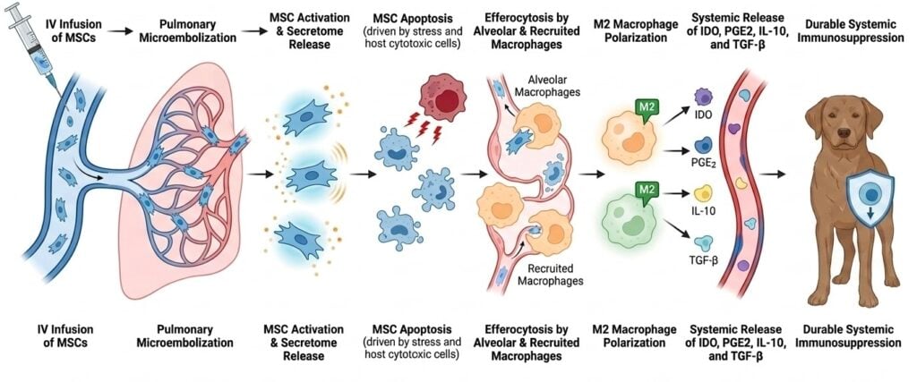
Once MSCs undergo apoptosis in the lungs, they expose phosphatidylserine on their outer membrane surface — the “eat-me” signal that attracts phagocytic cells. Alveolar macrophages and recruited monocytes engulf the apoptotic cells and their apoptotic bodies through the process of efferocytosis.16
What happens inside the macrophage after it engulfs an apoptotic MSC is where the real immune reprogramming occurs. The macrophages that phagocytose apoptotic MSCs are transcriptionally reprogrammed: they upregulate production of IDO (indoleamine 2,3-dioxygenase) and PGE2 (prostaglandin E2), two potent immunosuppressive mediators. 16 These reprogrammed macrophages shift from a pro-inflammatory M1 phenotype to an anti-inflammatory M2 phenotype and, critically, they continue producing immunosuppressive signals for days after the MSCs themselves are gone.
The apoptotic bodies released by dying MSCs carry specific proteins that appear to be the molecular signal for M2 macrophage polarization.17 Macrophage depletion experiments in multiple disease models have confirmed that abolishing macrophage function abolishes MSC therapeutic benefit entirely, establishing that macrophages are the final common effector of the entire pathway.
The cascade thus runs: IV infusion → pulmonary microembolization → MSC activation and secretome release → MSC apoptosis (driven by stress and host cytotoxic cells) → efferocytosis by alveolar and recruited macrophages → M2 macrophage polarization → systemic release of IDO, PGE2, IL-10, and TGF-β → durable systemic immunosuppression.
Every step of this cascade happens primarily in the lungs or is initiated there. This is why IV delivery is not a compromise — it is the route that most efficiently activates the mechanism.
CCL2: The Self-Amplifying Signal
One final piece completes the picture. It has been demonstrated in humans that apoptotic MSCs upregulate CCL2 (monocyte chemoattractant protein-1), which recruits additional monocytes from the bone marrow and circulation to the sites of apoptotic cell death.16 These incoming monocytes are in turn exposed to apoptotic MSC cargo and differentiate into additional M2-polarized anti-inflammatory macrophages. The initial wave of MSC apoptosis thus amplifies itself through a recruiting signal, creating a self-reinforcing anti-inflammatory cascade that extends well beyond the 24-hour window of MSC viability. This may be why therapeutic effects from a single infusion can persist for weeks to months.
What Does This Mean for Veterinary MSC Therapy?
The pulmonary first-pass effect was never the obstacle it was made out to be. Far from a dead end, the lung is where intravenously infused MSCs are activated, licensed, and ultimately cleared in a way that reprograms the immune system to do the therapeutic work – through secreted factors, apoptosis, and the macrophages that carry the signal forward. The lungs aren’t where IV stem cell therapy goes to fail; they’re where it goes to begin.
Key Takeaways
- Pulmonary retention after IV MSC administration is real and well documented.
- It limits the immediate passage of intact cells into systemic arterial circulation.
- It does not automatically mean therapeutic failure because MSC effects may also be mediated through secreted factors, apoptosis, efferocytosis, and recipient immune cells.
Frequently Asked Questions
What is the pulmonary first-pass effect?
The pulmonary first-pass effect describes the initial localization of intravenously administered cells in the pulmonary microvasculature before they enter systemic arterial circulation.
Why do mesenchymal stromal cells accumulate in the lungs?
MSC size and deformability relative to pulmonary capillaries are contributing factors. Receptor-mediated adhesion actively binds MSCs through a biologically mediated and advantageous process
Does pulmonary trapping mean IV stem cell therapy does not work?
No. It means that many stem cells do not immediately pass through the lungs. Therapeutic effects still occur through secreted mediators, immune-cell interactions, apoptosis, and efferocytosis. Some studies have also detected MSCs in other organs and injured tissues.
Do IV MSCs eventually leave the lungs?
Pulmonary signal generally decreases over time, and studies detect MSCs in other organs and injured tissues. The extent of viable-cell redistribution varies and depends heavily on the cell product, species, disease model, and detection method.
Why might MSC apoptosis be therapeutically relevant?
Experimental studies suggest that apoptotic MSCs can be engulfed by recipient macrophages and other phagocytes, helping shift these cells toward an immunoregulatory state.
References
- Fischer UM, Harting MT, Jimenez F, et al. Pulmonary Passage is a Major Obstacle for Intravenous Stem Cell Delivery: The Pulmonary First-Pass Effect. Stem Cells and Development. 2009;18(5):683-692. doi:10.1089/scd.2008.0253
- Sanchez-Diaz M, Quiñones-Vico MI, Sanabria de la Torre R, et al. Biodistribution of Mesenchymal Stromal Cells after Administration in Animal Models and Humans: A Systematic Review. J Clin Med. 2021;10(13):2925. doi:10.3390/jcm10132925
- Arzi B, Clark KC, Sundaram A, et al. Therapeutic Efficacy of Fresh, Allogeneic Mesenchymal Stem Cells for Severe Refractory Feline Chronic Gingivostomatitis. Stem Cells Translational Medicine. 2017;6(8):8. doi:10.1002/sctm.17-0035
- Beerts C, Brondeel C, Pauwelyn G, et al. Scintigraphic tracking of 99mTechnetium-labelled equine peripheral blood-derived mesenchymal stem cells after intravenous, intramuscular, and subcutaneous injection in healthy dogs. Stem Cell Res Ther. 2021;12(1):393. doi:10.1186/s13287-021-02457-9
- Kraitchman DL, Tatsumi M, Gilson WD, et al. Dynamic Imaging of Allogeneic Mesenchymal Stem Cells Trafficking to Myocardial Infarction. Circulation. 2005;112(10):1451-1461. doi:10.1161/CIRCULATIONAHA.105.537480
- Ferrini E, Stellari FF, Franceschi V, et al. Persistency of Mesenchymal Stromal/Stem Cells in Lungs. Front Cell Dev Biol. 2021;9:709225. doi:10.3389/fcell.2021.709225
- Spriet M, Hunt GB, Walker NJ, Borjesson DL. SCINTIGRAPHIC TRACKING OF MESENCHYMAL STEM CELLS AFTER PORTAL, SYSTEMIC INTRAVENOUS AND SPLENIC ADMINISTRATION IN HEALTHY BEAGLE DOGS. Vet Radiology Ultrasound. 2015;56(3):327-334. doi:10.1111/vru.12243
- Chow L, Johnson V, Coy J, Regan D, Dow S. Mechanisms of Immune Suppression Utilized by Canine Adipose and Bone Marrow-Derived Mesenchymal Stem Cells. Stem Cells and Development. 2017;26(5):374-389. doi:10.1089/scd.2016.0207
- Krampera M. Mesenchymal stromal cell ‘licensing’: a multistep process. Leukemia. 2011;25(9):1408-1414. doi:10.1038/leu.2011.108
- Lee RH, Pulin AA, Seo MJ, et al. Intravenous hMSCs Improve Myocardial Infarction in Mice because Cells Embolized in Lung Are Activated to Secrete the Anti-inflammatory Protein TSG-6. Cell Stem Cell. 2009;5(1):54-63. doi:10.1016/j.stem.2009.05.003
- Caplan AI, Correa D. The MSC: An Injury Drugstore. Cell Stem Cell. 2011;9(1):11-15. doi:10.1016/j.stem.2011.06.008
- Beerts C, Broeckx SY, Depuydt E, et al. Low-dose xenogeneic mesenchymal stem cells target canine osteoarthritis through systemic immunomodulation and homing. Arthritis Res Ther. 2023;25(1):190. doi:10.1186/s13075-023-03168-7
- Beerts C, Pauwelyn G, Depuydt E, et al. Homing of radiolabelled xenogeneic equine peripheral blood-derived MSCs towards a joint lesion in a dog. Front Vet Sci. 2022;9:1035175. doi:10.3389/fvets.2022.1035175
- Pang SHM, D’Rozario J, Mendonca S, et al. Mesenchymal stromal cell apoptosis is required for their therapeutic function. Nat Commun. 2021;12(1):6495. doi:10.1038/s41467-021-26834-3
- Galleu A, Riffo-Vasquez Y, Trento C, et al. Apoptosis in mesenchymal stromal cells induces in vivo recipient-mediated immunomodulation. Sci Transl Med. 2017;9(416):eaam7828. doi:10.1126/scitranslmed.aam7828
- Cheung TS, Giacomini C, Cereda M, et al. Apoptosis in mesenchymal stromal cells activates an immunosuppressive secretome predicting clinical response in Crohn’s disease. Molecular Therapy. 2023;31(12):3531-3544. doi:10.1016/j.ymthe.2023.10.004
- Kholodenko IV, Kholodenko RV, Majouga AG, Yarygin KN. Apoptotic MSCs and MSC-Derived Apoptotic Bodies as New Therapeutic Tools. CIMB. 2022;44(11):5153-5172. doi:10.3390/cimb44110351

Vice President of Veterinary Affairs at Gallant
Dr. Rebecca Windsor, DVM, DACVIM, is a board-certified veterinary neurologist with over 20 years of clinical experience and a strong record of scientific publication. She joined Gallant in 2025 and serves as Vice President of Veterinary Affairs.
Dr. Windsor specializes in veterinary regenerative medicine, with a focus on advancing FDA-approved, off-the-shelf mesenchymal stem cell therapies for pets. She develops educational platforms that translate the science, safety, and clinical efficacy of stem cell therapy for veterinary professionals. Since 2019, she has served as a Clinician Scientist at Ethos Discovery, where she leads the neurology research portfolio.