Canine Osteoarthritis Educational Series 2: Developmental Osteoarthritis Starts Earlier Than You Think
When we picture a dog with osteoarthritis (OA), many of us picture an older Labrador struggling to get up after laying on a slick floor. Unfortunately, for many of those dogs, arthritis has been active for many years, and we are seeing the accumulation of that chronic damage.
We now understand that most dogs do not develop arthritis in old age. For many, the disease begins during growth when the joints develop with improper fit and suboptimal physical function.
When a joint is not properly aligned or its cartilage surface is no longer smooth, the forces generated during movement become concentrated in small areas instead of being distributed evenly. This repetitive stress causes microtrauma, inflammation of the joint lining, cartilage damage, and changes to the underlying bone.1–3 Over time, abnormal biomechanics and chronic inflammation worsen, creating a progressive cycle that culminates in osteoarthritis.
In a recent study of dogs between 8 months and 4 years of age, nearly 40% had radiographic evidence of osteoarthritis in at least one appendicular joint, yet many showed only subtle clinical signs and received no specific treatment for OA.4 These findings reinforce an important concept: developmental orthopedic disease should be viewed as the beginning of a lifelong OA continuum rather than an isolated orthopedic diagnosis.
The greatest opportunity to influence long-term outcome of dogs with developmental arthritis occurs during the ‘golden window’ of growth from 4 months to 1.5 years old, when early diagnosis, appropriate surgery, rehabilitation, weight management, exercise modification, and multimodal medical care including regenerative therapies can reduce ongoing joint injury before irreversible changes occur.
What are the Most Common Reasons Young Dogs have Developmental Arthritis?
Developmental conditions account for the majority of OA that begins during growth and include hip dysplasia, elbow dysplasia, patellar luxation osteochondrosis/osteochondritis dissecans (OCD), and angular limb deformity (ALD). Although each arises from a different developmental abnormality, they all converge on the same endpoint: progressive cartilage damage, synovitis, and osteoarthritis.
Hip Dysplasia: Instability Creates Chronic Joint Trauma
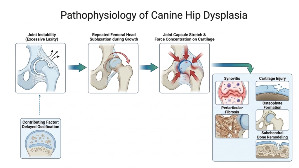
Canine hip dysplasia is fundamentally a disease of joint instability. Excessive laxity allows repeated subluxation of the femoral head during growth, stretching the joint capsule and concentrating forces on regions of cartilage not designed to bear load.5,6 This initiates synovitis, cartilage injury, periarticular fibrosis, osteophyte formation, and subchondral bone remodeling. Asynchronous and delayed ossification are major contributors of hip dysplasia.6,7
How Common Is Hip Dysplasia?
The frequency of hip dysplasia varies widely by breed, geography, and screening population. In a radiographic study of 27,710 dogs from 10 breeds, prevalence ranged from 5% in Siberian Huskies to 51.9% in Cane Corsos.8 In a study of more than 4000 large breed dogs from four specific breeds, passive hip laxity (which quantifies how far the femoral head can be displaced from the acetabulum) was the strongest measured risk factor for the development of hip OA.9 PennHIP radiographs are the most accurate method for canine hip radiograph positioning and can be performed as early as 16 weeks of age.10 Hip laxity measured in young dogs is associated with later OA risk. While PennHIP radiographs are the most specific for documenting laxity, regular hip extended views are often useful even in young dogs. OFA hip extended views to certify dogs as having good or excellent hips cannot be performed until they are two years old.
What are the Clinical Signs of Hip Dysplasia?
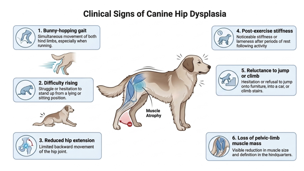
Clinical signs are classically biphasic. Juvenile dogs may experience pain associated with hip laxity, capsular inflammation, tearing, and microfracture of the dorsal acetabular rim. As periarticular fibrosis improves joint stability, clinical signs often diminish, creating the false impression that the problem has resolved. In reality, the disease continues to progress, and secondary osteoarthritis typically becomes clinically apparent later in life. Signs may then become less obvious as periarticular fibrosis improves stability, only to become prominent again later as secondary OA advances. Current understanding suggests that joint disease continues along a spectrum, even when the dog appears clinically better.6 Signs include bunny-hopping, difficulty rising, reduced hip extension, post-exercise stiffness, reluctance to jump or climb, and loss of pelvic-limb muscle. Even if patients undergo surgery, lifelong OA management remains important.
Why Early Recognition of Hip Dysplasia Matters
A puppy may run, play, and appear comfortable even while hip laxity is placing abnormal stress on the developing joint. Discuss screening and referral well before the 16-week visit whenever breed, family history, conformation, or physical examination raises concern. When PennHIP screening identifies a puppy with hip laxity, that puppy may be a candidate for a minimally invasive juvenile pubic symphysiodesis (JPS) surgery. This surgery involves cauterization of the pubic cartilage and has been shown to reduce the risk of hip OA in dogs with mild to moderate hip dysplasia.5 Dogs with suspect radiographs should be referred for orthopedic evaluation as soon as possible as the best outcomes are achieved when surgery is performed at approximately 15-20 weeks of age.5,7 This means veterinarians need to start conversations about lifelong joint health early, especially for large-breed dogs and breeds at increased risk. The 12-week puppy visit provides an ideal opportunity to discuss screening and early intervention while puppies are still within the optimal treatment window.
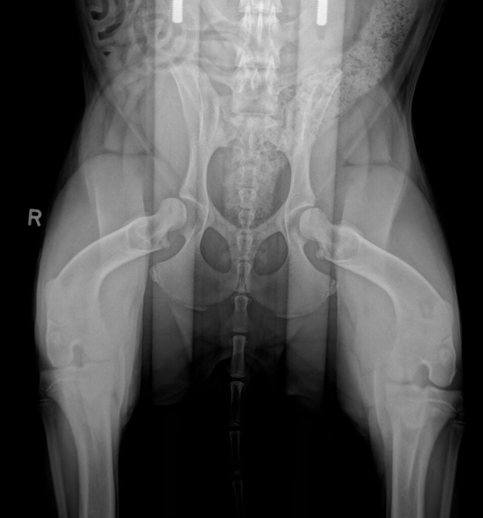
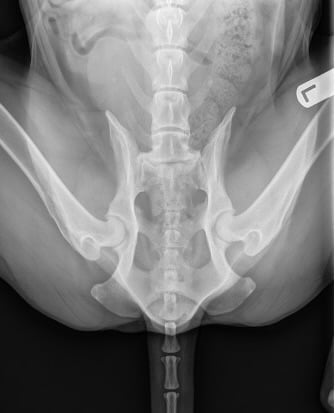
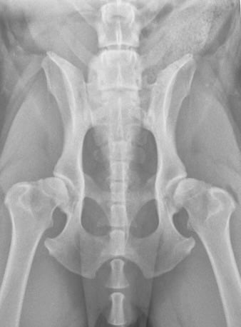
COMING SOON IN CANINE OSTEOARTHRITIS EDUCATIONAL SERIES 7: Screening Radiographs by Life Stage
We will review a radiographic screening protocol and provide more details on how to detect early signs of hip OA in radiographs. The results of these radiographs can be truly life-changing and guide recommendations for surgical referral, nutrition, supplements, regenerative medicine and rehabilitation for life.
Elbow Dysplasia: Several Disorders, One Progressive OA Pathway
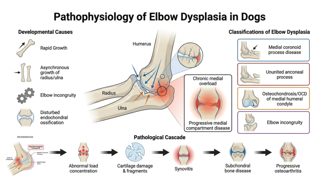
During rapid growth, asynchronous development of the radius and ulna, elbow incongruity, or disturbed endochondral ossification can alter the fit of the developing elbow and concentrate force on immature cartilage and subchondral bone.11
Elbow dysplasia includes four standard developmental classifications:
- Medial coronoid process disease
- Ununited anconeal process
- Osteochondrosis or osteochondritis dissecans of the medial humeral condyle
- Elbow incongruity
Although the initiating lesion differs among the various forms of elbow dysplasia, each disrupts the normal relationship between the radius, ulna, and humerus. Even subtle joint incongruity can concentrate forces on small areas of cartilage, leading to cartilage damage, fragment formation, subchondral bone disease, synovitis, and progressive osteoarthritis.11 Chronic medial overload, most commonly associated with medial coronoid process disease and elbow incongruity, may eventually produce extensive cartilage loss across the medial humeroulnar articulation, known as progressive medial compartment disease.12
How Common is Elbow Dysplasia?
In a 2024 radiographic-screening study of 17,861 dogs from 13 breeds, elbow dysplasia affected 11.4% overall, with breed-specific prevalence ranging from 1.1% to 32.2%.13
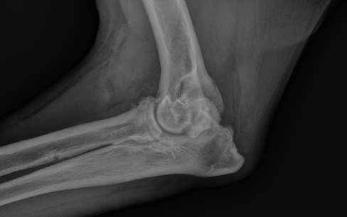
What are the Clinical Signs of Elbow Dysplasia?
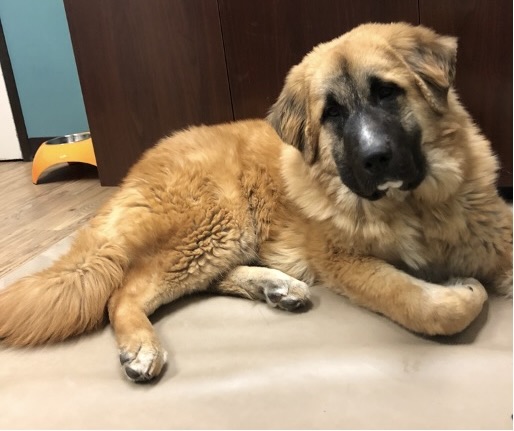
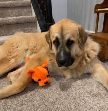
Young dogs often present with intermittent forelimb lameness, reduced elbow extension, stiffness after exercise, or subtle gait changes between 6-12 months of age. Another telling sign is outward rotation of the paw.
Why Early Recognition of Elbow Dysplasia Matters
Advanced imaging is often recommended in young dogs to better assess for elbow dysplasia. CT provides a more detailed assessment of bone alignment, elbow congruity, subchondral sclerosis, and medial coronoid pathology that may be missed or obscured on routine radiographs. Arthroscopy allows direct visualization of the articular cartilage and other intra-articular structures while also providing the opportunity to diagnose and treat selected lesions during the same procedure.11,14,15
Surgery may remove fragmented bone, stabilize or remove an ununited anconeal process, treat an OCD lesion, or improve load distribution within the joint, but even successful surgeries do not typically restore the joint to normal. Elbow dysplasia results in lifelong mechanical and inflammatory stress within the joint, often requiring ongoing reassessment, rehabilitation, weight management, medication, and individualized activity modification. Regenerative therapies, including platelet-rich plasma (PRP) and mesenchymal stromal cells (MSCs), may improve pain, mobility, and function and can serve as valuable adjuncts to surgery and long-term osteoarthritis management.16–19
Patellar Luxation: Abnormal Tracking Reshapes the Growing Limb
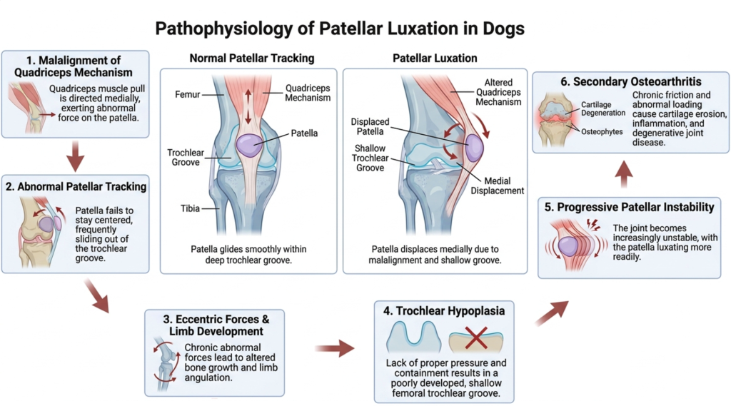
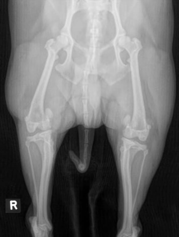
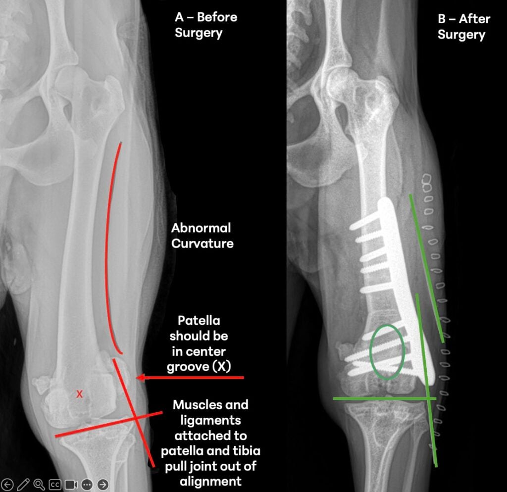
Patellar luxation occurs when the patella moves outside its normal track within the femoral trochlear groove. During growth, malalignment of the quadriceps mechanism results in abnormal patellar tracking. The resulting eccentric forces alter normal limb development, contributing to femoral and tibial angulation or rotation, trochlear hypoplasia, progressive patellar instability, and ultimately secondary osteoarthritis.20
How Common Is Patellar Luxation?
Patellar luxation was diagnosed in 1.3% of more than 210,000 dogs attending primary-care practices in England, with substantially higher prevalence among predisposed small and toy breeds.21
The patella may luxate medially (toward the inside of the limb) or laterally (toward the outside). In a large Swedish dog population, approximately 90% of cases were medial, while only 5.9% were lateral. Medial patellar luxation was most common in small breeds, whereas lateral luxation occurred more frequently in several larger breeds, particularly the Clumber Spaniel, Irish Wolfhound, and Coton de Tulear.22
What are the Clinical Signs of Patellar Luxation?
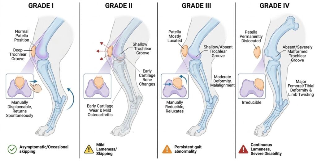
Clinical signs often include intermittent skipping, brief episodes of non-weight-bearing lameness, an abnormal sitting posture, reluctance to jump, or bowed and externally rotated pelvic limbs. Owners may report that their dog has “always moved that way,” leading these early signs to be dismissed as normal.
Patellar luxation is graded from I to IV based on how easily the patella luxates, whether it remains displaced, and whether it can be manually reduced into the trochlear groove. Although the grade describes the severity of patellar instability, treatment decisions should also consider the dog’s clinical signs, pain, gait abnormalities, cartilage damage, limb alignment, age, and evidence of disease progression, not the luxation grade alone.20,23
Why Early Recognition of Patellar Luxation Matters
Repeated patellar luxation alters normal patellofemoral contact, resulting in progressive cartilage injury and secondary osteoarthritis. In a study of surgically treated dogs, the severity of cartilage erosion was associated with higher luxation grade, increasing age, longer duration of clinical signs, and greater body weight.23
Early recognition provides an opportunity to intervene before irreversible cartilage damage occurs. Rather than waiting for lameness to become persistent or more severe, clinicians should recognize that worsening clinical signs often reflect cumulative cartilage injury and the progression of osteoarthritis, not simply advancement to a higher luxation grade.
Identifying patellar luxation during growth also allows for a multimodal treatment approach. Surgical correction, when indicated, can improve patellar tracking and limb alignment, while structured rehabilitation helps restore strength, function, and normal movement patterns. In a multicenter study of 400 dogs involving 574 surgically treated stifles, 88% had a good outcome.24In dogs with concurrent synovitis or osteoarthritis, regenerative therapies may serve as a valuable adjunct to reduce inflammation and improve joint function.20,23,25
Dogs with grade 2-4 patellar luxation should be referred for a surgical consultation between 3 and 6 months of age or as soon as the diagnosis is established. The goal is to address changes such as bowing of the femur and tibia during the early growth phase. In some cases, minimal surgical interventions to address these mechanical abnormalities early can make a big difference in the long-term. Early intervention reduces the risk of the predictable cascade of degenerative joint changes, pain, and muscle atrophy. Patellar luxation is also associated with increased risk of cruciate ligament tear, which was documented in 6.2% of dogs with patellar luxation compared with 0.67% of matched controls.22
Osteochondrosis and Osteochondritis Dissecans (OCD)
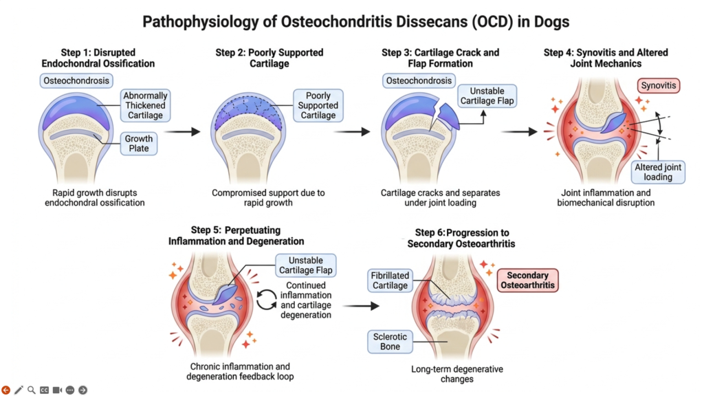
Osteochondrosis develops when endochondral ossification is disrupted during rapid growth, leaving a focal area of abnormally thickened, poorly supported cartilage. As the joint is loaded during normal activity, the weakened cartilage may crack and separate from the underlying bone, forming an unstable cartilage flap. This initiates synovitis, alters normal joint loading, causes pain, and begins the cascade toward secondary osteoarthritis.26
OCD most commonly affects the shoulder but can also occur in the elbow, stifle, and tarsus. Long-term prognosis and the risk of osteoarthritis depend on several factors, including the affected joint, lesion size, whether both limbs are involved, and how early the lesion is recognized and treated.11,26
How Common is OCD?
In a study of more than 600,000 dogs, appendicular osteochondrosis occurred at an incidence of approximately 4 cases per 10,000 dogs each year. Certain large and giant breeds showed significantly increased risk. The median age at diagnosis was approximately 9 months for appendicular osteochondrosis as a whole. Stifle lesions were diagnosed later.27
What are the Clinical Signs of OCD?
OCD typically presents during the first year of life as intermittent or progressive lameness that becomes more obvious after exercise. Shoulder lesions may cause pain with extension, a shortened forelimb stride, and shoulder-muscle asymmetry. Tarsal lesions cause pelvic limb lameness, visible tarsocrural swelling, and reduced joint motion. Lesions may be bilateral even when only one limb appears painful.26,27
The Importance of Early Recognition of OCD
An OCD lesion represents more than an unstable cartilage flap. It indicates that normal cartilage development and joint mechanics have already been disrupted during growth. Arthroscopy allows removal of unstable cartilage while providing direct assessment of the remaining articular surface. In a recent, randomized clinical trial, all dogs that completed the 1.5-year follow-up were sound and pain-free on joint palpation after arthroscopic treatment. However, CT still demonstrated osteoarthritic changes in 18 of 25 treated shoulders, highlighting that excellent clinical outcomes do not necessarily indicate restoration of a normal joint.28
Evidence supporting the use of platelet-rich plasma (PRP) or mesenchymal stromal cells (MSCs) specifically following canine OCD surgery remains limited. However, based on the broader canine OA literature, these therapies may be considered as adjuncts for dogs with persistent synovitis, pain, or secondary osteoarthritis after the primary lesion has been addressed.
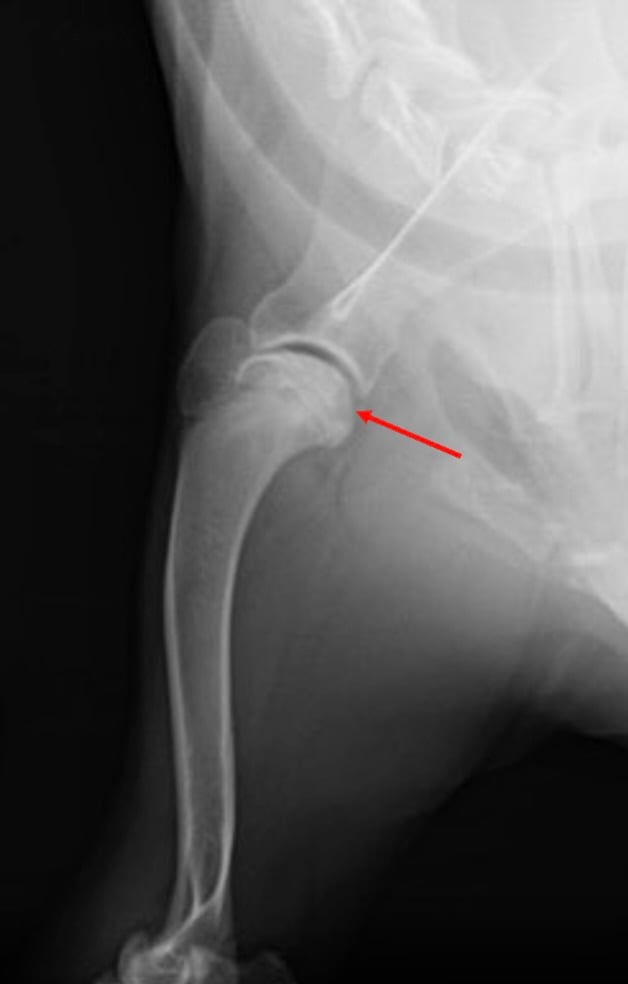
Angular Limb Deformity: When Uneven Growth Changes the Whole Limb
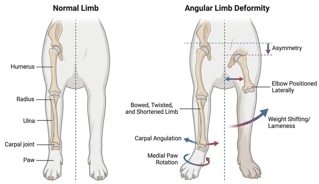
Angular limb deformity (ALD) develops when inherited bone growth or premature, asymmetric closure of a growth plate causes paired bones to grow at different rates. In the forelimb, early closure of the distal ulnar growth plate can tether the still-growing radius, contributing to valgus, rotation, procurvatum, radial bowing, elbow incongruity, and limb shortening.29,30
As alignment changes, forces are redistributed through the elbow, carpus, shoulder, surrounding soft tissues, and opposite limb.
Which Dogs Are Most Often Affected?
A reliable general-population prevalence has not been established. Breed-associated ALD is documented in short-limbed, chondrodysplastic breeds including the Dachshund, Skye Terrier, and Glen of Imaal Terrier.31 Acquired ALD can occur in any growing dog after premature or asymmetric physeal closure, including following growth-plate trauma.30,31 The risk for OA development in ALD affected dogs is unknown. In one cross-sectional breed specific study, OA was present in 35.2% of Skye Terrier elbows and 6.7% of Dachshund elbows. Greater elbow incongruity and lateral radial-head subluxation were associated with OA.31
What are the Clinical Signs of Angular Limb Deformity?
Angular limb deformity may appear as:
- A paw that turns inward or outward
- A carpus that angles to one side
- An elbow positioned farther away from the chest
- A bowed, twisted, or shortened limb
- Uneven paw placement
- Progressive asymmetry, lameness, or weight shifting
A breed-typical limb shape may be common without being biomechanically neutral.
Why Early Recognition of Angular Limb Deformity Matters
In a four-center referral study of juvenile dogs treated for premature distal ulnar physeal closure, patients ranged from 4 to 10.8 months of age and presented with visible carpal valgus and varying degrees of forelimb lameness. Distal ulnar ostectomy helped halt progression in appropriately selected growing dogs, but it did not reverse established angulation during the study period.29
Refer as soon as progressive angulation or asymmetric growth is recognized and ideally while meaningful growth remains. Early imaging and orthopedic referral provide the best opportunity to slow disease progression, improve joint congruity, and perform corrective surgery before irreversible secondary joint damage develops.29,30
Conclusion
Developmental orthopedic diseases may have different underlying causes, but they all share the same consequence: abnormal joint mechanics that initiate a lifelong cycle of cartilage injury, inflammation, and progressive osteoarthritis. While many affected dogs remain active and outwardly comfortable during growth, the disease process is already underway long before chronic pain becomes apparent.
Recognizing these conditions early provides an opportunity to change the course of disease. Appropriate screening, timely referral, surgical intervention when indicated, rehabilitation, weight management, exercise modification, and multimodal medical therapy can all help reduce ongoing joint damage and preserve long-term mobility. Although surgery can improve joint mechanics, it cannot restore an already injured joint to normal, making lifelong osteoarthritis management an essential part of patient care.
The ultimate goal is not simply to correct a developmental abnormality, it is to preserve healthy joint function for as many years as possible. By recognizing developmental orthopedic disease as the beginning of the osteoarthritis continuum, veterinarians can intervene earlier, set realistic expectations for caregivers, and help dogs maintain comfort, mobility, and quality of life throughout their lives.
Coming up next in the Canine Osteoarthritis Educational Series: Understanding the Pathophysiology of Cruciate Ligament Disease
References
1. Anderson KL, Zulch H, O’Neill DG, Meeson RL, Collins LM. Risk Factors for Canine Osteoarthritis and Its Predisposing Arthropathies: A Systematic Review. Front Vet Sci. 2020;7:220. doi:10.3389/fvets.2020.00220
2. Marcellin-Little DJ, Hulse DA, Huntingford JL, et al. A proposed framework for practical multimodal management of osteoarthritis in growing dogs. Front Vet Sci. 2025;12:1565922. doi:10.3389/fvets.2025.1565922
3. Loeser RF, Goldring SR, Scanzello CR, Goldring MB. Osteoarthritis: A disease of the joint as an organ. Arthritis & Rheumatism. 2012;64(6):1697-1707. doi:10.1002/art.34453
4. Enomoto M, De Castro N, Hash J, et al. Prevalence of radiographic appendicular osteoarthritis and associated clinical signs in young dogs. Sci Rep. 2024;14(1):2827. doi:10.1038/s41598-024-52324-9
5. Linn KA. Juvenile Pubic Symphysiodesis. Veterinary Clinics of North America: Small Animal Practice. 2017;47(4):851-863. doi:10.1016/j.cvsm.2017.03.004
6. King MD. Etiopathogenesis of Canine Hip Dysplasia, Prevalence, and Genetics. Veterinary Clinics of North America: Small Animal Practice. 2017;47(4):753-767. doi:10.1016/j.cvsm.2017.03.001
7. Lopez M, Schachner E. Diagnosis, prevention, and management of canine hip dysplasia: a review. VMRR. Published online May 2015:181. doi:10.2147/VMRR.S53266
8. Baldinger A, Genevois JP, Moissonnier P, et al. Prevalence of canine hip dysplasia in 10 breeds in France, a retrospective study of the 1997-2017 radiographic screening period. Clegg S, ed. PLoS ONE. 2020;15(7):e0235847. doi:10.1371/journal.pone.0235847
9. Runge JJ, Kelly SP, Gregor TP, Kotwal S, Smith GK. Distraction index as a risk factor for osteoarthritis associated with hip dysplasia in four large dog breeds*. J of Small Animal Practice. 2010;51(5):264-269. doi:10.1111/j.1748-5827.2010.00937.x
10. Patricelli AJ, Dueland RT, Adams WM, Fialkowski JP, Linn KA, Nordheim EV. Juvenile Pubic Symphysiodesis in Dysplastic Puppies at 15 and 20 Weeks of Age. Veterinary Surgery. 2002;31(5):435-444. doi:10.1053/jvet.2002.34766
11. Vezzoni A, Benjamino K. Canine Elbow Dysplasia. Veterinary Clinics of North America: Small Animal Practice. 2021;51(2):439-474. doi:10.1016/j.cvsm.2020.12.007
12. Bruecker KA, Benjamino K, Vezzoni A, et al. Canine Elbow Dysplasia. Veterinary Clinics of North America: Small Animal Practice. 2021;51(2):475-515. doi:10.1016/j.cvsm.2020.12.008
13. Roels J, Genevois JP, Fostier-Humbert M, et al. Prevalence of elbow dysplasia in 13 dog breeds in France: a retrospective radiographic study (2002–2022). ajvr. 2024;85(6):ajvr.23.12.0290. doi:10.2460/ajvr.23.12.0290
14. Kähn H, Zablotski Y, Meyer-Lindenberg A. Therapeutic success in fragmented coronoid process disease and other canine medial elbow compartment pathology: a systematic review with meta-analyses. Front Vet Sci. 2023;10:1228497. doi:10.3389/fvets.2023.1228497
15. Burton NJ. Review of minimally invasive surgical procedures for assessment and treatment of medial coronoid process disease. Veterinary Surgery. 2023;52(6):790-800. doi:10.1111/vsu.13986
16. Matos Cruz AM, Mason DR. Owner assessed outcomes following elbow arthroscopy with or without platelet rich plasma for fragmented medial coronoid process. Front Vet Sci. 2022;9:938706. doi:10.3389/fvets.2022.938706
17. Black LL, Gaynor J, Adams C, et al. Effect of intraarticular injection of autologous adipose-derived mesenchymal stem and regenerative cells on clinical signs of chronic osteoarthritis of the elbow joint in dogs. Vet Ther. 2008;9(3):3.
18. Kim SE, Pozzi A, Yeh J chern, et al. Intra-Articular Umbilical Cord Derived Mesenchymal Stem Cell Therapy for Chronic Elbow Osteoarthritis in Dogs: A Double-Blinded, Placebo-Controlled Clinical Trial. Front Vet Sci. 2019;6:474. doi:10.3389/fvets.2019.00474
19. Olsen A, Johnson V, Webb T, Santangelo K, Dow S, Duerr F. Evaluation of Intravenously Delivered Allogeneic Mesenchymal Stem Cells for Treatment of Elbow Osteoarthritis in Dogs: A Pilot Study. Vet Comp Orthop Traumatol. 2019;32(03):03. doi:10.1055/s-0039-1678547
20. Perry KL, Déjardin LM. Canine medial patellar luxation. J of Small Animal Practice. 2021;62(5):315-335. doi:10.1111/jsap.13311
21. O’Neill DG, Meeson RL, Sheridan A, Church DB, Brodbelt DC. The epidemiology of patellar luxation in dogs attending primary-care veterinary practices in England. Canine Genet Epidemiol. 2016;3(1):4. doi:10.1186/s40575-016-0034-0
22. Engdahl K, Bergström A, Höglund O, Hanson J. The epidemiology of patellar luxation in an insured Swedish dog population. Preventive Veterinary Medicine. 2023;220:106034. doi:10.1016/j.prevetmed.2023.106034
23. Kim HW, Kim YS, Kim WK, Kang KW, Kang BJ. Medial patellar luxation induces cartilage erosion in dogs: a retrospective study of prevalence and risk factors. ajvr. 2024;85(11):ajvr.24.07.0190. doi:10.2460/ajvr.24.07.0190
24. Bosio F, Bufalari A, Peirone B, Petazzoni M, Vezzoni A. Prevalence, treatment and outcome of patellar luxation in dogs in Italy: A retrospective multicentric study (2009–2014). Vet Comp Orthop Traumatol. 2017;30(05):364-370. doi:10.3415/VCOT-16-05-0073
25. Brondeel C, Pauwelyn G, De Bakker E, Saunders J, Samoy Y, Spaas JH. Review: Mesenchymal Stem Cell Therapy in Canine Osteoarthritis Research: “Experientia Docet” (Experience Will Teach Us). Front Vet Sci. 2021;8:668881. doi:10.3389/fvets.2021.668881
26. Stokes R, Dycus D. The Shoulder Joint and Common Abnormalities. Veterinary Clinics of North America: Small Animal Practice. 2021;51(2):323-341. doi:10.1016/j.cvsm.2020.11.002
27. Engdahl K, Höglund O, Hedhammar Å, Hanson J, Bergström A. The epidemiology of osteochondrosis in an insured Swedish dog population. Preventive Veterinary Medicine. 2024;228:106229. doi:10.1016/j.prevetmed.2024.106229
28. Heikkilä H, Reunanen V, Hyytiäinen HK, Junnila JJT, Laitinen-Vapaavuori O, Keränen P. Randomized, Blinded, Controlled Clinical Trial of Polylactide–Collagen Scaffold in Treatment of Shoulder Osteochondritis Dissecans in Dogs. Vet Comp Orthop Traumatol. 2024;37(06):286-296. doi:10.1055/s-0044-1788726
29. Christopher S. Short term outcomes and complications of distal ulnar ostectomy in 23 juvenile dogs with carpal valgus secondary to discordant radial-ulnar physeal growth. Front Vet Sci. 2022;9:971527. doi:10.3389/fvets.2022.971527
30. Fox DB. Physeal Injuries and Angular Limb Deformities. Veterinary Clinics of North America: Small Animal Practice. 2021;51(2):305-322. doi:10.1016/j.cvsm.2020.11.003 31. Lappalainen AK, Pulkkinen HSM, Mölsä S, Junnila J, Hyytiäinen HK, Laitinen-Vapaavuori O. Breed-typical front limb angular deformity is associated with clinical findings in three chondrodysplastic dog breeds. Front Vet Sci. 2023;9:1099903. doi:10.3389/fvets.2022.1099903

Vice President of Veterinary Affairs at Gallant
Dr. Rebecca Windsor, DVM, DACVIM, is a board-certified veterinary neurologist with over 20 years of clinical experience and a strong record of scientific publication. She joined Gallant in 2025 and serves as Vice President of Veterinary Affairs.
Dr. Windsor specializes in veterinary regenerative medicine, with a focus on advancing FDA-approved, off-the-shelf mesenchymal stem cell therapies for pets. She develops educational platforms that translate the science, safety, and clinical efficacy of stem cell therapy for veterinary professionals. Since 2019, she has served as a Clinician Scientist at Ethos Discovery, where she leads the neurology research portfolio.