Canine Osteoarthritis Educational Series 3: The Inflammatory Nature of Cranial Cruciate Ligament Disease in Dogs
Cranial cruciate ligament (CCL) disease often begins well before complete ligament rupture. Recognizing early changes including synovitis, altered joint mechanics, muscle loss, and subtle instability gives us more opportunities to protect the meniscus, preserve function, and manage secondary osteoarthritis (OA) throughout the patient’s life.
Consider a young Rottweiler that presents after a weekend of play with intermittent hind-limb lameness. She is still weight-bearing, and the stifle is mildly effusive, but there is no convincing cranial drawer. After several days of an NSAID, she looks almost normal. That improvement matters, relieving pain and improving comfort are important treatment goals. However, improvement in comfort does not necessarily mean the underlying disease process has stopped. Clinical signs of OA can wax and wane even as synovitis, abnormal joint loading, muscle loss, and tissue damage continue.
CCL rupture can occur acutely after substantial trauma; however, in dogs, CCL rupture more commonly represents the later stage of progressive ligament degeneration. As the ligament weakens, it becomes increasingly vulnerable to repetitive normal loading or relatively minor injury.1 Multiple factors contribute to CCL disease development and progression in dogs, including genetic susceptibility, body weight and conformation, chronic synovial inflammation, and ligament degeneration, 1–4. Importantly, synovitis and early CCL fiber disruption may be present before the stifle becomes clinically unstable. In one study, all clinically stable contralateral stifles evaluated arthroscopically had synovitis, and 75% already had CCL fiber disruption.5 In other words, the dramatic “tear” may be the event we notice, not the event that started the disease.
This changes how we think about secondary OA. Rather than viewing CCL rupture as a single orthopedic event, we can take a 360-degree, lifetime approach to joint health and identify disease as early as possible, control synovitis and pain, preserve muscle and movement, protect the meniscus, restore joint mechanics when indicated, and continue OA management long after the initial injury. Surgery can be an important point along that trajectory, but it is neither the beginning nor the end of joint care.
The Normal Stifle at a Glance
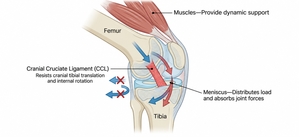
In a healthy stifle, stability depends on multiple structures working together. The CCL limits cranial movement and internal rotation of the tibia, while the menisci distribute load and absorb forces between the femur and tibia. Surrounding muscles provide dynamic support. Together, these structures help keep the stifle stable and allow forces to move through the joint in a controlled manner.6
Which Dogs Are at Higher Risk for CCL Disease?
CCL disease is common; however, risk is not evenly distributed across the canine population. Genetics set the stage, while conformation, body condition, age, and patellar stability help shape an individual dog’s risk.
In a UK primary-care population of more than 170,000 dogs, 0.56% had a recorded diagnosis of CCL disease. Rottweilers, Golden Retrievers, West Highland White Terriers, Yorkshire Terriers, and Staffordshire Bull Terriers were at increased odds for diagnosis. Studies in Labrador Retrievers support a polygenic susceptibility to CCL disease.2 Age, body condition, limb and stifle conformation, muscle function, and patellar stability can all influence joint mechanics and may contribute to an individual dog’s risk. 1,2,7
For our young Rottweiler, the combination of breed predisposition, recurrent effusion, and intermittent lameness should raise our index of suspicion. Rather than waiting for obvious instability to develop, we can compare both stifles carefully, assess thigh muscle mass and patellar stability, and consider imaging when the results are likely to change management.
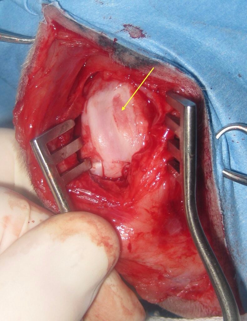
For more on how conformation and developmental orthopedic disease can set the stage for secondary OA, read Gallant’s Canine Osteoarthritis Educational Series 2: Developmental Osteoarthritis Starts Earlier Than You Think.
What Happens When the CCL Starts to Fail?
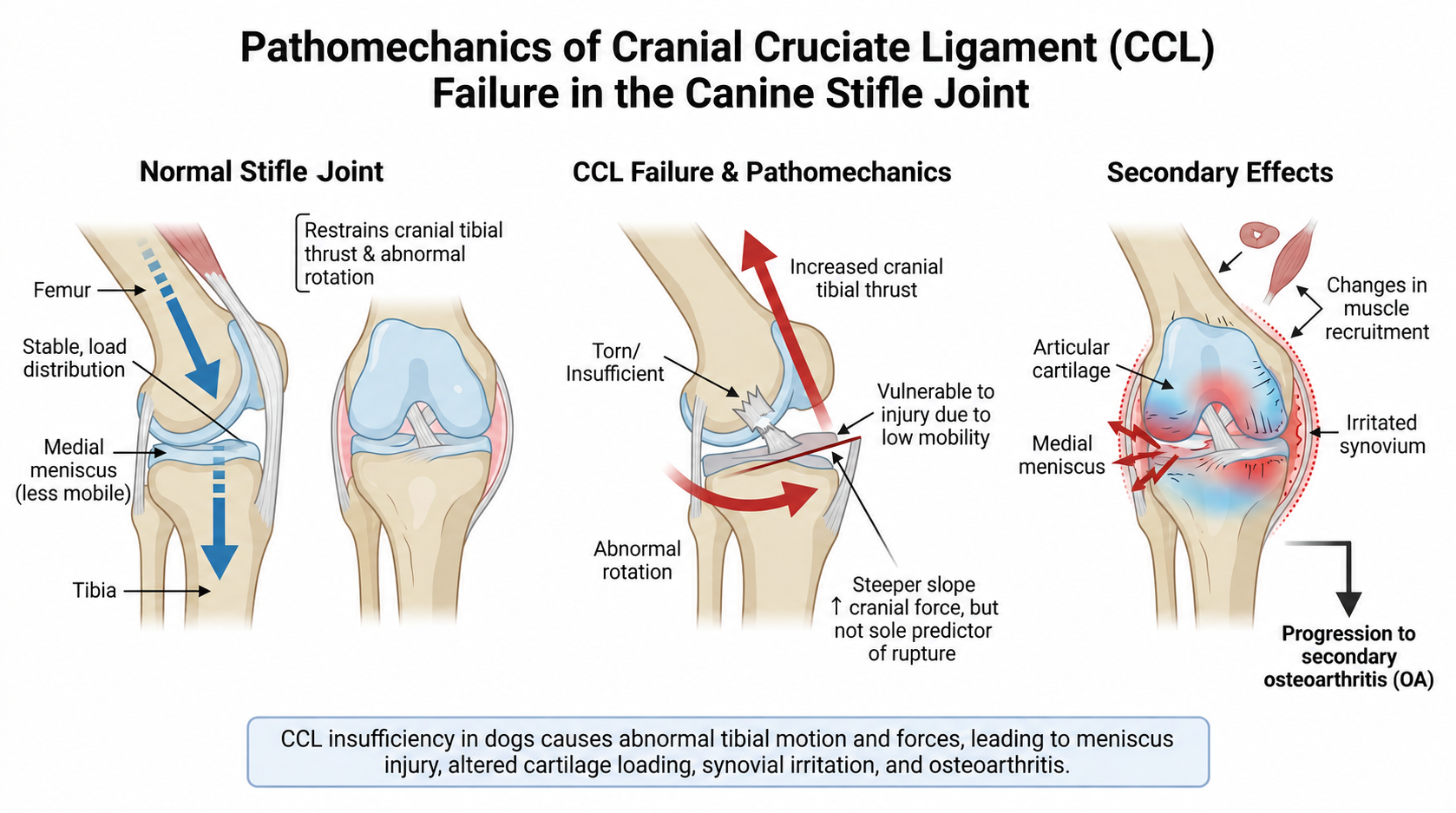
The medial meniscus is less mobile than the lateral meniscus. Under normal conditions, this relative stability helps it manage load, but when the CCL becomes insufficient, it also makes the medial meniscus particularly vulnerable to injury.
During weight bearing, compression across the sloped tibial plateau generates cranial tibial thrust. An intact CCL restrains this motion. As the CCL loses function, the tibia increasingly thrusts cranially and rotates abnormally beneath the femur, altering how forces are distributed across the articular cartilage and menisci.6
A steeper tibial plateau can increase the cranially directed component of this force, although tibial plateau angle alone does not predict whether CCL rupture will occur.1,6
As stability deteriorates, the consequences extend beyond the ligament itself. The meniscus is exposed to abnormal shear forces, cartilage experiences uneven loading, muscle recruitment changes, and the synovium is repeatedly irritated. These altered mechanics help drive the development and progression of secondary OA.
What Is Happening Biologically Inside the Stifle?
Mechanical instability is only part of the disease process. CCL disease also involves an active inflammatory and immune response within the joint. Synovial tissue from affected dogs contains macrophages, T and B lymphocytes, plasma cells, and antigen-presenting cells; antibodies directed against collagen have also been identified.1,3 These findings do not establish CCL disease as a classic autoimmune disorder, but they demonstrate that ligament degeneration occurs alongside an immune response that may contribute to persistent synovitis.
Proinflammatory macrophage activity is present in both partial and complete CCL disease.4 Inflammatory mediators, including IL-1β, TNF-α, IL-6, and IL-17, promote cartilage-catabolic signaling, while chemokines recruit additional inflammatory cells into the joint. Downstream enzymes, including matrix metalloproteinases (MMPs) and ADAMTS aggrecanases, degrade components of the cartilage extracellular matrix.1,10
As cartilage breaks down, matrix fragments and biomarkers such as CTX-II, C2C, aggrecan/proteoglycan fragments, and glycosaminoglycan fragments can be detected in synovial fluid and, for some markers, blood or urine.11–13
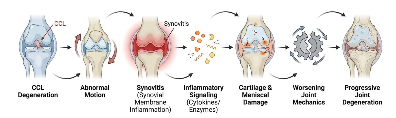
The result is a self-perpetuating interaction between mechanics and inflammation:
CCL degeneration → abnormal joint motion → synovitis → inflammatory and catabolic signaling → cartilage and meniscal damage → worsening joint mechanics
Importantly, greater arthroscopic synovitis has been associated with longer duration of clinical signs and more advanced cartilage damage, reinforcing that inflammation is an important component of CCL-associated OA progression, not simply a consequence of instability.14
How Do We Know What Stage of CCL Disease We’re Treating?
We perform diagnostics to define what is happening in the stifle and determine whether the joint remains functionally stable.
Once CCL disease is suspected, the next question is: where is this patient along the disease continuum?
A thorough orthopedic examination including comparison of both pelvic limbs, assessment of muscle symmetry, evaluation of joint effusion, and stability testing combined with appropriate imaging can help distinguish early or partial CCL disease from a functionally unstable tear. This assessment can also identify concurrent OA, patellar disease, and evidence of meniscal injury.
Radiographs provide valuable information about joint effusion, osteophyte formation, subchondral bone changes, patellar abnormalities, and established OA. Importantly, radiographic abnormalities may be present even before obvious clinical instability develops. In one study, 34.5% of palpably normal contralateral stifles in dogs with unilateral CCL rupture already had an abnormal infrapatellar fat-pad sign on radiographs.15
Musculoskeletal ultrasound can provide additional information about joint effusion, synovial thickening, CCL fiber integrity, and meniscal pathology. In the hands of a trained sonographer, musculoskeletal ultrasound is also useful in evaluating the meniscus. In one study comparing ultrasonography with arthroscopy, ultrasound identified medial meniscal lesions with 95% sensitivity and 82% specificity, although diagnostic accuracy may vary with operator experience.16
Together, these findings help define not simply whether CCL disease is present, but what stage of disease we are treating, information that can guide both immediate management and longer-term joint care.
Why Timing Matters: Protect the Meniscus
The treatment priority changes when a partial, functionally stable injury progresses to mechanical instability.
In a study of 443 CCL-deficient stifles, 36.3% had medial meniscal tears. Complete CCL rupture increased the odds of meniscal injury nearly 13-fold, and the risk increased by approximately 2.6% for each additional week of lameness.17 Meniscal injury has also been associated with poorer objective weight bearing and a greater OA burden.18,19
Functionally Stable Partial Tear
We cannot reliably prevent every partially damaged CCL from eventually progressing to complete rupture. However, earlier recognition gives us an opportunity to address many of the consequences that are already developing, including synovitis, pain, muscle loss, asymmetrical loading, excess body weight, and altered movement patterns.
Anti-inflammatory therapy can help control pain and synovitis, while rehabilitation can help preserve muscle mass, range of motion, neuromuscular control, and more symmetrical limb loading.20,21 Evidence supports therapeutic exercise as part of rehabilitation following CCL surgery, although study quality and specific rehabilitation protocols vary.
Earlier recognition may also provide an opportunity to consider regenerative medicine as part of a multimodal joint-preservation strategy. Canine studies suggest that mesenchymal stromal cell (MSC)-based therapies can modulate inflammatory signaling within the joint, although their ability to prevent complete CCL rupture or halt OA progression has not been established.22,23 Additional studies are needed and ongoing.
Complete, Unstable Tear
Once functional stability is lost, the treatment priority changes. Each loading cycle exposes the meniscus and cartilage to abnormal translation and rotation.
For an appropriate surgical candidate, timely stabilization becomes the priority, with the goal of restoring a more favorable mechanical environment and limiting continued exposure of the meniscus and cartilage to abnormal joint motion.17
For many families, the cost and emotional weight of surgery can make an initial trial of pain medications and rest appealing. The concern is that improvement in pain does not correct mechanical instability. In an unstable stifle, joint damage may continue during observation: meniscal risk increases with longer duration of lameness, synovitis is associated with disease duration and cartilage damage, and OA changes can develop rapidly.14,17
If surgery cannot be pursued immediately or while a functionally stable partial tear is being monitored, that period can still be used proactively. Controlling synovitis, maintaining an appropriate body condition, preserving muscle and movement through rehabilitation, and considering appropriate joint-directed or regenerative therapies can all remain part of the management plan.20–23
Importantly, joint care does not end with stabilization. Surgery addresses the mechanical instability, but it does not eliminate the underlying OA process. Continued attention to inflammation, muscle strength, movement, body condition, and long-term joint health remains important after surgery.
Why Is Surgery a Critical Pivot Point?
For dogs with mechanically unstable CCL disease, surgical stabilization can substantially improve limb function and create a more favorable mechanical environment within the stifle.24,25 Several surgical approaches are available, including osteotomy-based procedures such as TPLO and TTA, extracapsular stabilization techniques, and, less commonly, intracapsular or reconstructive procedures. Comparing the indications and outcomes of these techniques is beyond the scope of this article.
Importantly, surgery does not reconstruct or replace the damaged CCL. Instead, it alters the geometry of the proximal tibia to reduce the cranially directed shear forces that would normally be restrained by an intact CCL. This allows the CCL-deficient stifle to achieve functional stability during weight bearing.
Surgery therefore represents an important pivot point in the CCL disease continuum: the mechanical problem may be substantially improved, but the biologic consequences of joint disease do not simply reset. Synovitis, cartilage injury, meniscal pathology, muscle loss, and established OA may still require ongoing management after stabilization. Radiographic OA can continue to progress following surgery and other stabilization procedures, reinforcing the need to view surgery as one component of lifelong joint care rather than the endpoint of treatment.
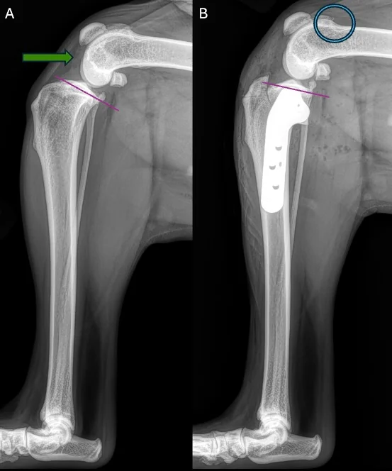
What surgery does not do is eliminate established synovitis, restore damaged cartilage, or reverse existing OA. Dogs can regain excellent weight-bearing function after TPLO even as radiographic OA continues to progress over time.26 The healed-TPLO image in Figure 5B illustrates this important distinction. The surgically stabilized stifle has a substantially greater radiographic OA burden than the contralateral stifle. Although a single radiograph cannot tell us when those changes developed or how quickly they progressed, it reinforces an important concept: successful mechanical stabilization and control of the underlying OA process are distinct treatment goals.
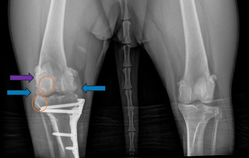
Objective gait studies demonstrate that limb function can continue to improve for several months after surgery, with near-normal function often occurring later in the recovery period.27 If recovery plateaus, lameness recurs, or function remains meaningfully limited at approximately 4 months, this provides a practical opportunity to assess for meniscal disease, muscle deficits, or another source of pain.16,20,21,26,27
Same Surgery. Different Stifles.
The same mechanical diagnosis can present with very different inflammatory and functional disease burdens.
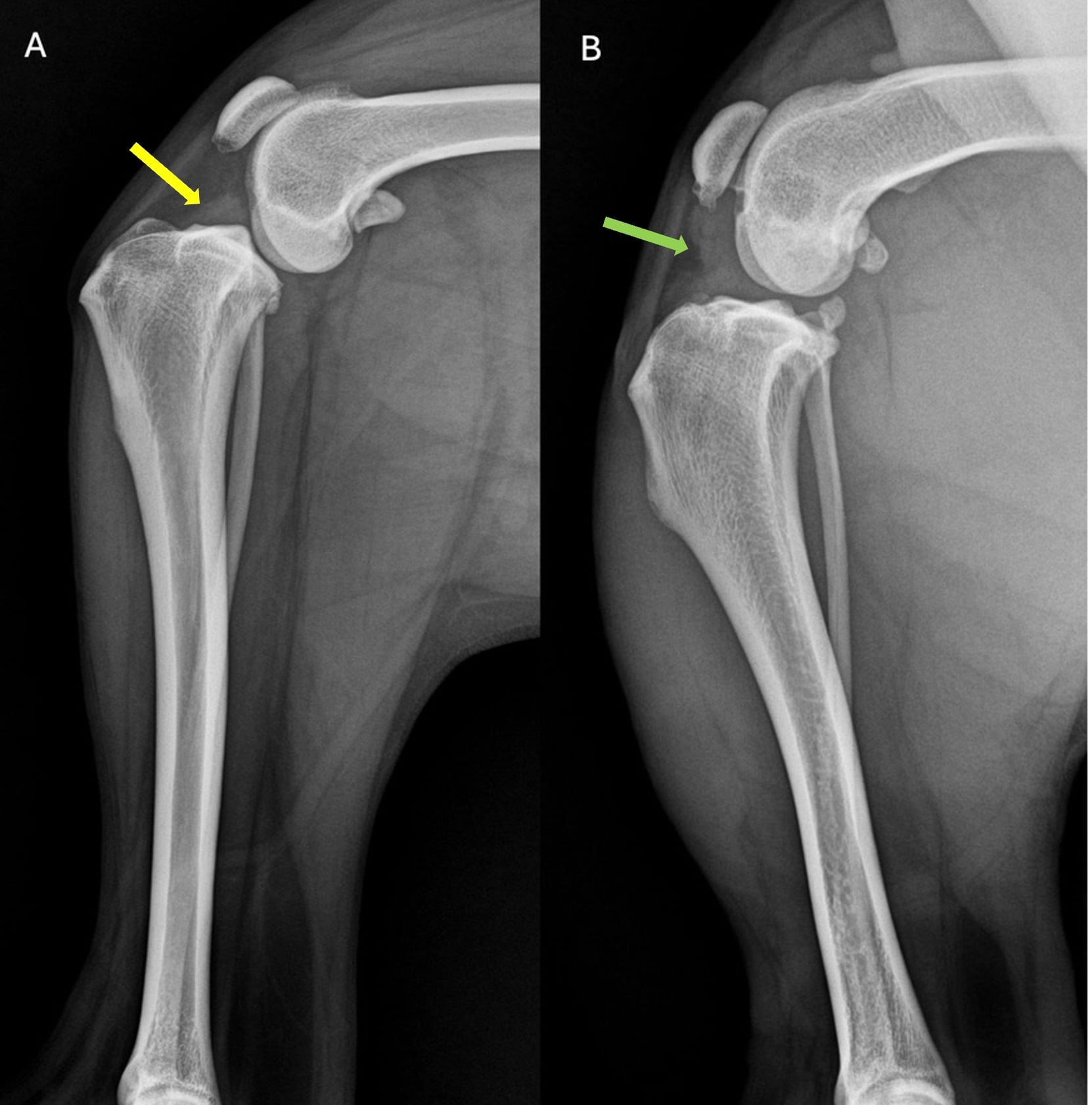
Consider two 2-year-old dogs with complete CCL tears. Both require surgical stabilization, but they arrive at surgery with very different stifles and different needs beyond the surgery itself.
Patient A (Figure 7A) had minimal joint effusion and synovitis but substantial muscle atrophy. For this patient, restoring muscle mass, strength, and limb symmetry may be an important priority alongside stabilization.
Patient B (Figure 7B) had marked synovitis and joint effusion. Here, greater emphasis may be needed on controlling the inflammatory joint environment before and after surgery.
These differences matter. Greater arthroscopic synovitis has been associated with more advanced cartilage lesions,14 while muscle loss can contribute to persistent functional impairment and requires targeted rehabilitation.
Surgery addresses mechanical instability, but it does not address every component of CCL disease. The broader treatment plan should reflect the inflammatory, structural, and functional needs of the individual patient, addressing the whole joint and whole patient.
Putting it Together: Best Care Practice for Canine CCL Disease
The best approach to CCL disease involves matching treatment to the factors driving disease at each stage. Early management may focus on controlling synovitis, preserving muscle and movement, optimizing body condition, and closely monitoring stability. If the stifle becomes mechanically unstable, stabilization becomes a priority to restore more normal mechanics and reduce ongoing risk to the meniscus and cartilage. Rehabilitation helps rebuild physical capacity, while long-term OA management continues to address inflammation, pain, muscle health, and mobility.20,21,24–26
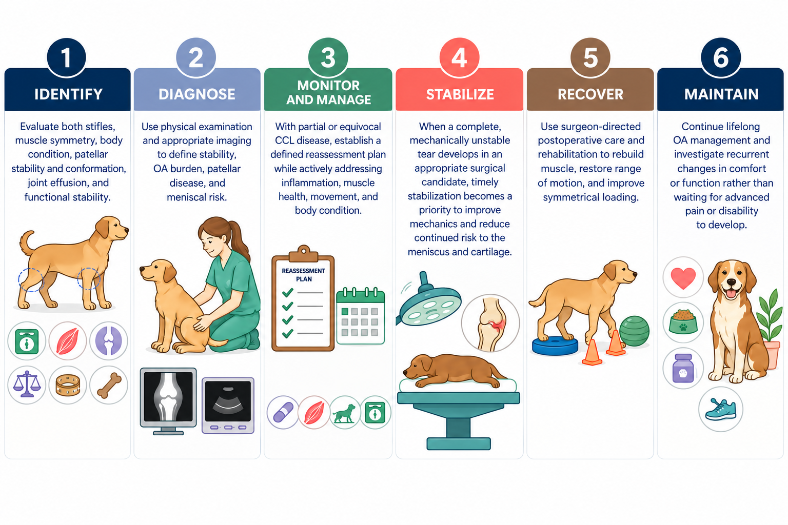
Figure 8. Cranial Cruciate Ligament Disease Management Timeline
Key Takeaways
- Earlier recognition creates more opportunities to intervene. Synovitis, altered loading, muscle loss, and cartilage injury may begin before complete CCL rupture or obvious instability.
- Improvement in comfort does not mean the underlying disease process has stopped. Clinical signs can improve while inflammation and structural disease continue.
- Protect the meniscus. As CCL insufficiency progresses to complete instability, the risk of meniscal injury increases substantially.
- Diagnostics guide the next decision. The key distinction is whether the stifle remains functionally stable enough for active management and monitoring or has become unstable enough that restoring mechanics should become the priority.
- Surgery treats instability, not the entire OA process. Long-term care should continue to address inflammation, muscle health, movement, body condition, and pain.
- Recovery deserves reassessment. Persistent or recurrent functional limitations around 4 months after stifle surgery—particularly TPLO—provide a practical reason to investigate residual joint, meniscal, muscle, or other pain-related problems rather than assuming that additional time is the only answer.
References
1. Niebauer GW, Restucci B. Etiopathogenesis of Canine Cruciate Ligament Disease: A Scoping Review. Animals. 2023;13(2):187. doi:10.3390/ani13020187
2. Cook SR, Conzemius MG, McCue ME, Ekenstedt KJ. SNP‐based heritability and genetic architecture of cranial cruciate ligament rupture in Labrador Retrievers. Animal Genetics. 2020;51(5):824-828. doi:10.1111/age.12978
3. Doom M, De Bruin T, De Rooster H, Van Bree H, Cox E. Immunopathological mechanisms in dogs with rupture of the cranial cruciate ligament. Veterinary Immunology and Immunopathology. 2008;125(1-2):143-161. doi:10.1016/j.vetimm.2008.05.023
4. Yarnall BW, Chamberlain CS, Hao Z, Muir P. Proinflammatory polarization of stifle synovial macrophages in dogs with cruciate ligament rupture. Veterinary Surgery. 2019;48(6):1005-1012. doi:10.1111/vsu.13261
5. Bleedorn JA, Greuel EN, Manley PA, et al. Synovitis in Dogs with Stable Stifle Joints and Incipient Cranial Cruciate Ligament Rupture: A Cross‐Sectional Study. Veterinary Surgery. 2011;40(5):531-543. doi:10.1111/j.1532-950X.2011.00841.x
6. Spinella G, Arcamone G, Valentini S. Cranial Cruciate Ligament Rupture in Dogs: Review on Biomechanics, Etiopathogenetic Factors and Rehabilitation. Veterinary Sciences. 2021;8(9):186. doi:10.3390/vetsci8090186
7. Taylor‐Brown FE, Meeson RL, Brodbelt DC, et al. Epidemiology of Cranial Cruciate Ligament Disease Diagnosis in Dogs Attending Primary‐Care Veterinary Practices in England. Veterinary Surgery. 2015;44(6):777-783. doi:10.1111/vsu.12349
8. Hewicker-Trautwein M, Carter SD, Bennett D, Kelly DF. Immunocytochemical demonstration of lymphocyte subsets and MHC class II antigen expression in synovial membranes from dogs with rheumatoid arthritis and degenerative joint disease. Veterinary Immunology and Immunopathology. 1999;67(4):341-357. doi:10.1016/S0165-2427(98)00229-3
9. Campbell CA, Horstman CL, Mason DR, Evans RB. Severity of patellar luxation and frequency of concomitant cranial cruciate ligament rupture in dogs: 162 cases (2004–2007). javma. 2010;236(8):887-891. doi:10.2460/javma.236.8.887
10. Malek S, Weng HY, Martinson SA, Rochat MC, Béraud R, Riley CB. Evaluation of serum MMP-2 and MMP-3, synovial fluid IL-8, MCP-1, and KC concentrations as biomarkers of stifle osteoarthritis associated with naturally occurring cranial cruciate ligament rupture in dogs. Zhang C, ed. PLoS ONE. 2020;15(11):e0242614. doi:10.1371/journal.pone.0242614
11. Prink A, Hayashi K, Kim SY, Kim J, Kapatkin A. Evaluation of a Collagenase Generated Osteoarthritis Biomarker in the Synovial Fluid from Elbow Joints of Dogs with Medial Coronoid Disease and Unaffected Dogs. Veterinary Surgery. 2010;39(1):65-70. doi:10.1111/j.1532-950X.2009.00604.x
12. Fujita Y, Hara Y, Nezu Y, Schulz KS, Tagawa M. Proinflammatory Cytokine Activities, Matrix Metalloproteinase‐3 Activity, and Sulfated Glycosaminoglycan Content in Synovial Fluid of Dogs with Naturally Acquired Cranial Cruciate Ligament Rupture. Veterinary Surgery. 2006;35(4):369-376. doi:10.1111/j.1532-950X.2006.00159.x
13. De Bakker E, Broeckx B, Demeyere K, Stroobants V, Van Ryssen B, Meyer E. Detection of osteoarthritis in dogs by metabolic, pro-inflammatory and degenerative synovial fluid biomarkers and traditional radiographic screening: A pilot study. Veterinary Immunology and Immunopathology. 2021;237:110252. doi:10.1016/j.vetimm.2021.110252
14. Lemmon EA, Xiao R, Mauck RL, Agnello KA. Arthroscopic synovitis severity scoring in canine stifles with cranial cruciate ligament disease. Veterinary Surgery. 2025;54(3):486-495. doi:10.1111/vsu.14222
15. Fuller MC, Hayashi K, Bruecker KA, et al. Evaluation of the radiographic infrapatellar fat pad sign of the contralateral stifle joint as a risk factor for subsequent contralateral cranial cruciate ligament rupture in dogs with unilateral rupture: 96 cases (2006–2007). javma. 2014;244(3):328-338. doi:10.2460/javma.244.3.328
16. Franklin SP, Cook JL, Cook CR, Shaikh LS, Clarke KM, Holmes SP. Comparison of ultrasonography and magnetic resonance imaging to arthroscopy for diagnosing medial meniscal lesions in dogs with cranial cruciate ligament deficiency. javma. 2017;251(1):71-79. doi:10.2460/javma.251.1.71
17. Hayes GM, Langley‐Hobbs SJ, Jeffery ND. Risk factors for medial meniscal injury in association with cranial cruciate ligament rupture. J of Small Animal Practice. 2010;51(12):630-634. doi:10.1111/j.1748-5827.2010.01003.x
18. Wustefeld‐Janssens BG, Pettitt RA, Cowderoy EC, et al. Peak Vertical Force and Vertical Impulse in Dogs With Cranial Cruciate Ligament Rupture and Meniscal Injury. Veterinary Surgery. 2016;45(1):60-65. doi:10.1111/vsu.12419
19. Fung C, Ficklin M, Okafor CC. Associations between meniscal tears and various degrees of osteoarthritis among dogs undergoing TPLO for cranial cruciate ligament rupture. BMC Res Notes. 2023;16(1):36. doi:10.1186/s13104-023-06307-0
20. Monk ML, Preston CA, McGowan CM. Effects of early intensive postoperative physiotherapy on limb function after tibial plateau leveling osteotomy in dogs with deficiency of the cranial cruciate ligament. American Journal of Veterinary Research. 2006;67(3):529-536. doi:10.2460/ajvr.67.3.529
21. Alvarez LX, Repac JA, Kirkby Shaw K, Compton N. Systematic review of postoperative rehabilitation interventions after cranial cruciate ligament surgery in dogs. Veterinary Surgery. 2022;51(2):233-243. doi:10.1111/vsu.13755
22. Canapp SO, Leasure CS, Cox C, Ibrahim V, Carr BJ. Partial Cranial Cruciate Ligament Tears Treated with Stem Cell and Platelet-Rich Plasma Combination Therapy in 36 Dogs: A Retrospective Study. Front Vet Sci. 2016;3. doi:10.3389/fvets.2016.00112
23. Muir P, Hans EC, Racette M, et al. Autologous Bone Marrow-Derived Mesenchymal Stem Cells Modulate Molecular Markers of Inflammation in Dogs with Cruciate Ligament Rupture. Goncalves R, ed. PLoS ONE. 2016;11(8):e0159095. doi:10.1371/journal.pone.0159095
24. Wemmers AC, Charalambous M, Harms O, Volk HA. Surgical treatment of cranial cruciate ligament disease in dogs using Tibial Plateau Leveling Osteotomy or Tibial Tuberosity Advancement–A systematic review with a meta-analytic approach. Front Vet Sci. 2022;9:1004637. doi:10.3389/fvets.2022.1004637
25. Gordon-Evans WJ, Griffon DJ, Bubb C, Knap KM, Sullivan M, Evans RB. Comparison of lateral fabellar suture and tibial plateau leveling osteotomy techniques for treatment of dogs with cranial cruciate ligament disease. javma. 2013;243(5):675-680. doi:10.2460/javma.243.5.675
26. Shimada M, Mizokami N, Ichinohe T, et al. Long-term outcome and progression of osteoarthritis in uncomplicated cases of cranial cruciate ligament rupture treated by tibial plateau leveling osteotomy in dogs. J Vet Med Sci. 2020;82(7):908-916. doi:10.1292/jvms.19-0613
27. Amimoto H, Koreeda T, Wada N. Evaluation of recovery of limb function by use of force plate gait analysis after tibial plateau leveling osteotomy for management of dogs with unilateral cranial cruciate ligament rupture. ajvr. 2019;80(5):461-468. doi:10.2460/ajvr.80.5.461

Dr. Marie Bartling is a veterinarian with more than 20 years of clinical experience specializing in integrative medicine, pain management, rehabilitation, and regenerative therapies.
Her approach combines conventional medicine with advanced therapies to manage complex medical conditions, musculoskeletal disease, and chronic pain. She is passionate about translating emerging science into practical, evidence-based strategies that improve patient health, mobility, and quality of life.

Vice President of Veterinary Affairs at Gallant
Dr. Rebecca Windsor, DVM, DACVIM, is a board-certified veterinary neurologist with over 20 years of clinical experience and a strong record of scientific publication. She joined Gallant in 2025 and serves as Vice President of Veterinary Affairs.
Dr. Windsor specializes in veterinary regenerative medicine, with a focus on advancing FDA-approved, off-the-shelf mesenchymal stromal cell (MSC) therapies for pets. She develops educational platforms that translate the science, safety, and clinical efficacy of stem cell therapy for veterinary professionals. Since 2019, she has served as a Clinician Scientist at Ethos Discovery, where she leads the neurology research portfolio.