Feline Osteoarthritis Series Part 2: Diagnosing Osteoarthritis in the Cat
In the first installment of this series we established that feline osteoarthritis (OA), also called degenerative joint disease (DJD), is one of the most common and under-recognized chronic feline diseases, affecting more than half of cats over six years of age and around 90% of geriatric cats.1–3 Despite this high prevalence, OA is commonly missed by veterinarians and caregivers, likely because the signs of OA in cats can be difficult to recognize. Unlike the obviously lame arthritic dog, arthritic cats often mask symptoms, part of an innate protective mechanism. Diagnosis rests on caregiver-reported behavioral change, the orthopedic examination, and diagnostic imaging.4–6 This installment of our feline osteoarthritis education series walks through each of those elements, the particular challenges of feline radiography, and the structured scoring tools that turn a vague clinical impression into a measurable, trackable diagnosis.
Why Diagnosing OA in Cats Is So Difficult
Cats are masters of concealment. As both predator and prey species, they instinctively hide signs of weakness, and overt lameness (the hallmark of canine OA) is the exception rather than the rule in cats.4,5 Two features of feline OA compound this. First, the disease is usually bilateral and frequently affects multiple joints, so there is no “good limb” for the cat to shift weight onto and no asymmetry for the observer to notice. Second, the joint changes are often biomechanically subtle. The result is a cat that does not limp but instead simply does less, easily misinterpreted as normal aging.
The veterinary clinic environment can exacerbate the diagnostic challenge. Quite often cats are examined on a table with little to no time to observe them walking around the room because they are fearful. When they do a move around, a fearful stressed cat often freezes, crouches, or struggles, and almost never moves as it would at home. The most valuable diagnostic information frequently comes from outside the clinic, relying heavily on the caregiver account and ability to video at-home gait. 7,8
Recognizing the Clinical Signs
Behavioral and Lifestyle Changes: The Cornerstone
Because lameness is uncommon, the practical diagnosis of feline OA begins with behavioral and lifestyle changes. Owner-observation studies and an expert behavioral consensus have identified a recognizable cluster of signs.7–9 Hesitancy or difficulty jumping or using stairs and change in gait are reported in at least three-quarters of cats with OA.7 When affected cats are treated with analgesic medications, many of these particular behaviors improve, supporting their value as genuine markers of joint pain rather than incidental aging.8
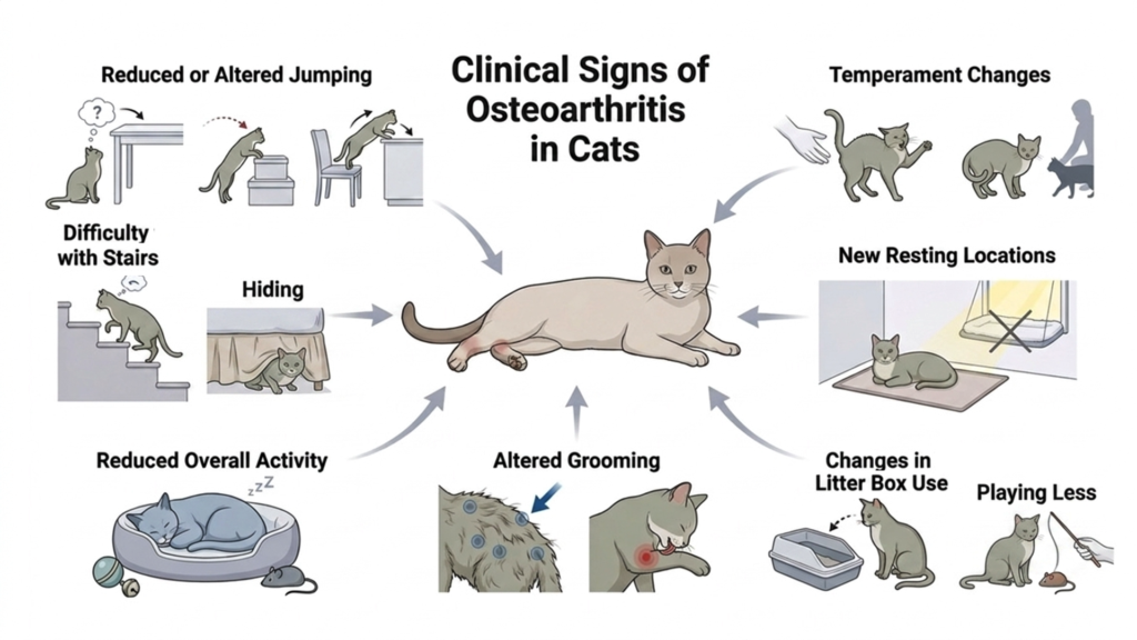
Commonly reported changes include:
- Reduced or altered jumping—hesitating before a jump, jumping to lower surfaces, jumping in stages, or no longer jumping up to favored high spots
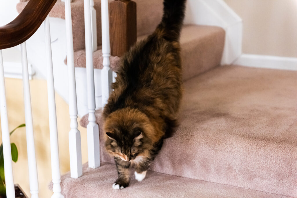
- Difficulty with stairs, or taking them slowly or one at a time
- Reduced overall activity, more sleeping, and less play or hunting
- Altered grooming – an unkempt or matted coat (especially over the back and hindquarters), or excessive licking/over-grooming directed at a painful joint
- Changes in litter box use, including eliminating just outside a high-sided box that has become difficult to climb into
- New resting locations chosen for accessibility rather than height or warmth
- Temperament change – defensive behavior, avoidance behavior, social withdrawal, protective response when being handled, petted, or lifted
No single sign is diagnostic, and many overlap with other diseases of older cats. Their power lies in the pattern and the change from an individual cat’s own baseline, which is precisely why a structured history is so important.9,10
The Orthopedic Examination and Its Limits
A careful hands-on examination remains essential and can yield real findings: pain or guarding on joint manipulation, reduced range of motion, periarticular thickening, crepitus, and muscle atrophy over chronically painful limbs. The joints most commonly affected are the elbow, hip, stifle, tarsus, and shoulder.2,3,11 However, a stressed cat may tense against any manipulation, masking focal pain, while a relaxed cat may tolerate handling that would otherwise be uncomfortable. Therefore, a normal-seeming examination does not rule out OA, and examination findings must be interpreted alongside the history, not in isolation.4
The Role and Limits of Radiography
What Feline OA Looks Like on Radiographs
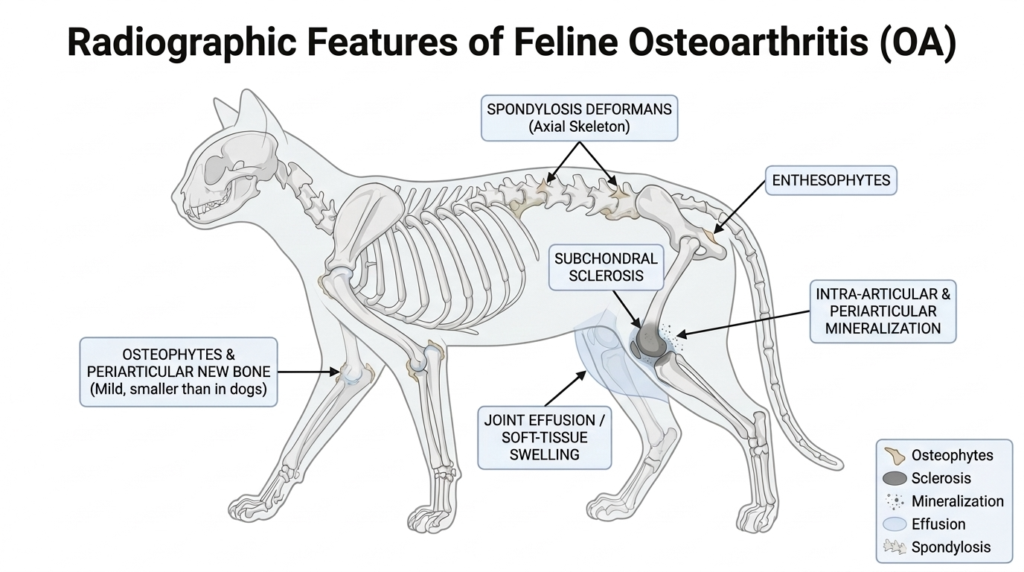
Radiography remains the most accessible imaging tool for confirming structural joint disease and is the basis for nearly all feline OA prevalence data.2,11–13 Recognized radiographic features of feline OA include osteophytes and periarticular new bone (typically milder than in dogs), enthesophytes, subchondral sclerosis, intra-articular and periarticular mineralization, joint effusion or soft-tissue swelling, and, in the axial skeleton, spondylosis deformans. In the overweight cat in particular, the stifle is a frequently affected and radiographically informative joint.14 In a cohort of 101 cats undergoing arthritis screening with orthogonal radiographs, the elbow was the most commonly affected appendicular joint, followed by the stifle; involvement was bilateral in roughly 91% of affected cats, and mineralization within the medial meniscus was present in nearly all affected stifles 11
The Challenges of Radiographic Diagnosis
Radiography in the cat carries important caveats. Because feline osteophytosis is often modest, early or mild changes are easily overlooked, and the diagnostic yield depends heavily on technique. In a study screening 101 cats, orthogonal imaging identified appendicular OA in 74% of cats and spondylosis in 41%; however, hip OA and stifle meniscal mineralization were detectable only on the ventrodorsal/cranio-caudal projection, while spondylosis was visible only on the lateral view; a single radiographic plane would therefore have missed clinically meaningful disease in many cats.11 Good positioning frequently requires sedation, which a painful or comorbid older cat may not tolerate well, and the bilateral nature of feline OA removes the contralateral “normal” limb that radiologists rely on for comparison.
The single most important caveat, however, is the well-documented discordance between radiographic severity and clinical signs. Cats with marked radiographic OA may behave near-normally, while cats with substantial pain-associated behavioral change may show only mild radiographic findings.2–4 In the 101 cat screening cohort described above, only a single owner reported lameness despite widespread radiographic disease, and appendicular OA was generally not painful on palpation, reinforcing that imaging findings routinely exceed what is detectable clinically.11 Radiographs document structure; they do not measure pain. The decision to treat should never rest on radiographs alone and should be based on the combination of clinical signs and our knowledge of disease progression, regardless of how severe the disease may or may not seem on a radiograph.
Why Owner Assessment and Home Video Are Essential
If the clinic environment makes gait assessment and examination challenging and radiographs do not correlate with pain, the home environment becomes the most reliable window into a cat’s mobility. Caregivers observe their cats daily, across the surfaces, jumps, and routines that arthritis makes more challenging. Studies comparing caregiver-perceived signs with veterinary findings show that caregivers detect meaningful, pain-relevant change that may not be apparent in the veterinary clinic.7,8
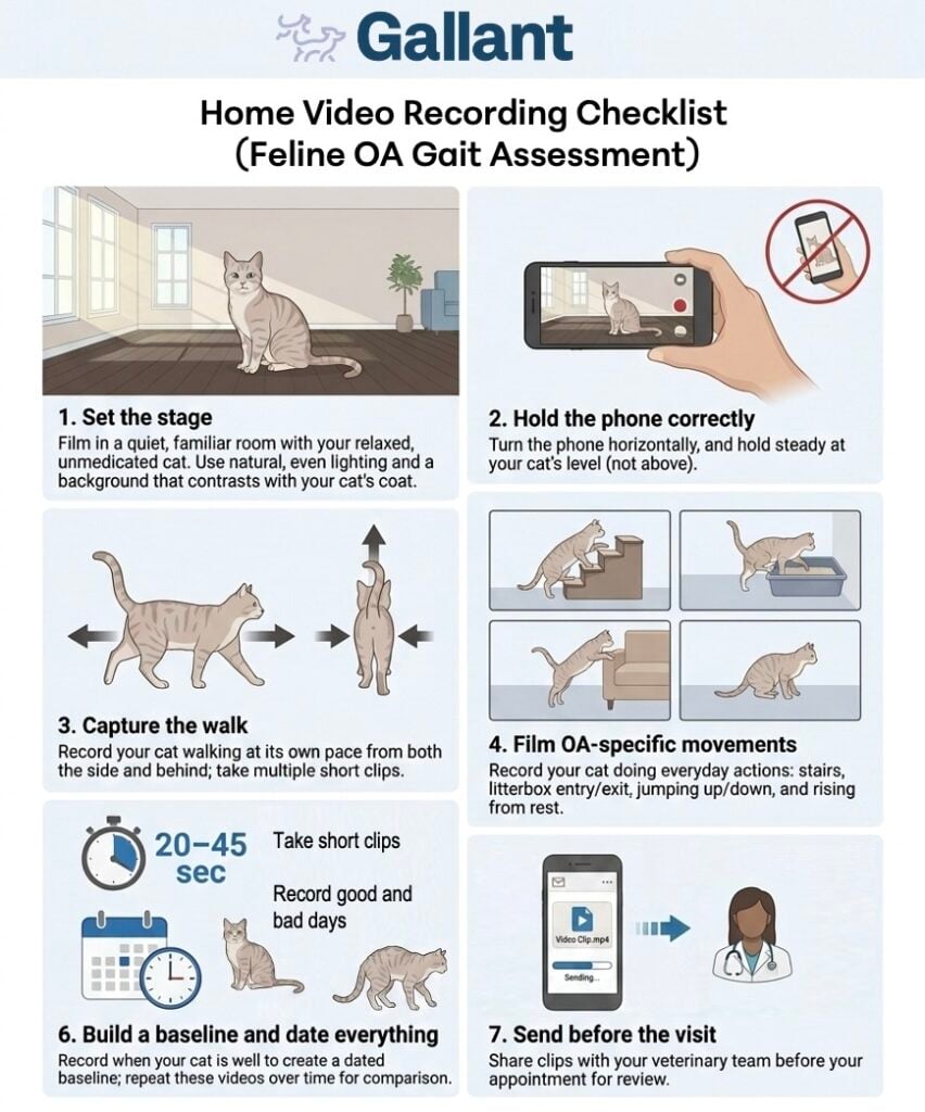
Home video has become a particularly powerful tool. Short clips of a cat negotiating stairs, jumping up to or down from furniture, walking along a hallway, or stepping in and out of the litter box capture natural movement that is simply unobtainable in the clinic. Asking owners to film these specific activities turns subjective impressions into reviewable evidence and helps track response to treatment over time. Researchers developing feline pain-assessment tools have specifically called for capturing home video before and after treatment to better characterize the behaviors that change with pain relief.15
Engaging our feline caregivers is critical to long-term management success. Living with a chronically painful cat carries a measurable emotional toll and expenditure of time and dollars, and this caregiver burden is increasingly recognized.16 Caregivers who understand what they are watching for become better observers and more committed partners in long-term management.
Scoring Tools: Clinical Metrology Instruments
The feline pain management community has developed structured, validated questionnaires and scales for caregivers and clinicians that convert behavioral observation into numbers, giving us a basis to measure and discuss comfort in cats. These tools are commonly referred to as clinical metrology instruments (CMIs). Their value is twofold: they prompt caregivers and clinicians to ask about the right behaviors, and they generate a repeatable baseline score that can be re-measured to judge treatment response.15,17
Caregiver-Completed Instruments
Caregiver-completed CMIs are the most critical component of feline OA assessment because cat owners regularly witness the cat’s natural behavior. The most comprehensive survey is the Feline Musculoskeletal Pain Index (FMPI), a questionnaire in which caregivers rate their cat’s ability to perform a series of everyday activities that has been compared to objective activity-monitor data in a double-masked, placebo-controlled crossover trial of 66 cats where the 17-item FMPI (scored as a percentage of the possible total) accurately detected the effect of analgesic treatment.15 A second validated owner survey, the Client-Specific Outcome Measures (CSOMs), take a complementary, individualized approach: the owner identifies three activities that are specifically difficult for that particular cat (i.e. jumping onto a bed, using stairs, getting in and out of the litterbox) and grades them at each visit.15,17 Validated questionnaires capture part of the picture and are best read alongside other evidence.6,10,15

In-Clinic Pain Scales
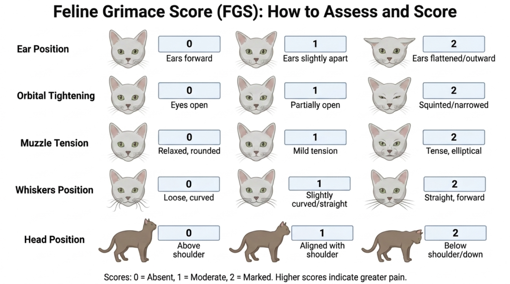
Because cats mask pain, we often use their facial expressions and posture to determine how comfortable they are. Structured behavioral observation tools have been developed for the veterinary team. The Feline Grimace scale (shown below) illustrates signs of discomfort based on whisker placement, eye and ear and head position.18,19 These instruments are best validated for acute pain, and while interest in applying facial and behavioral scoring to chronic OA pain is growing, they do not yet replace owner-completed CMIs for the longitudinal assessment of chronic feline OA.19
Activity Monitoring
Objective activity monitoring, called accelerometry, offers a sensor-based measure of how much a cat moves and has been used to detect treatment-related changes in cats with OA.15,17 An accelerometer is a small, lightweight motion sensor, similar in principle to the step-counter inside a smartphone or a fitness tracker. In cats it is usually mounted on the collar, where it continuously records movement and converts it into “activity counts”, a numerical measure of how much and how vigorously the cat is moving over time. Because the device runs quietly in the background in the cat’s own home, it captures normal day-to-day behavior over days to weeks rather than as a single snapshot in time. In a typical assessment, the cat wears the monitor for a baseline period of one to two weeks to establish the typical activity level and daily pattern before any treatment begins. The same monitor is then worn through the treatment period so that activity during therapy can be compared directly against that individual cat’s own baseline.17,20,21
Analgesic Trial
A pragmatic and widely used diagnostic strategy is the analgesic trial. When history, examination, and imaging point toward OA, a carefully chosen course of analgesia is given and the cat is reassessed, ideally using the same CMI and home observations recorded before treatment. Improvement in mobility and behavior supports the diagnosis.8
When interpreting response to therapy for any animal trial, we must be careful to account for the caregiver placebo effect. This can be exceptionally true for cats given how difficult it is to recognize clinical signs, and feline OA studies have demonstrated a sizeable caregiver placebo effect.15 Recognizing this phenomenon and defining what constitutes a clinically relevant degree of pain relief is central to interpreting CMIs and treatment trials honestly.15,22
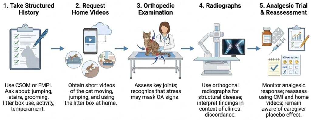
Because no single test is definitive, a comprehensive approach is recommended including the following:
- Take a structured history using a validated CMI such as the CSOM or FMPI, asking specifically about jumping, stairs, grooming, litter box use, activity, and temperament
- Request short home videos of the cat moving, jumping, and using the litter box in its normal environment
- Perform a careful orthopedic examination, focusing on the elbow, hip, stifle, tarsus, and shoulder, while recognizing that stress can mask findings
- Use targeted, orthogonal radiographs to document structural disease—while interpreting them in light of known radiographic–clinical discordance
- Consider a monitored analgesic trial, reassessing with the same CMI and home observations and remaining mindful of the caregiver placebo effect
As we evolve our knowledge and medical care for cats with osteoarthritis, we are finding that we have even more opportunities to improve the quality of life and mobility for cats who have long lived under the radar of our clinical attention.
Once OA is recognized, the focus shifts to treatment. The next installment, Feline Osteoarthritis Educational Series 3: Traditional Treatment Options for OA in the Cat, will review the established, evidence-based strategies for managing feline OA pain and improving quality of life.
References
- Hardie EM, Roe SC, Martin FR. Radiographic evidence of degenerative joint disease in geriatric cats: 100 cases (1994–1997). J Am Vet Med Assoc. 2002;220(5):628-632. doi:10.2460/javma.2002.220.628
- Lascelles BDX, Henry III JB, Brown J, et al. Cross-Sectional Study of the Prevalence of Radiographic Degenerative Joint Disease in Domesticated Cats. Vet Surg. 2010;39(5):535-544. doi:10.1111/j.1532-950X.2010.00708.x
- Slingerland LI, Hazewinkel HAW, Meij BP, Picavet Ph, Voorhout G. Cross-sectional study of the prevalence and clinical features of osteoarthritis in 100 cats. Vet J. 2011;187(3):304-309. doi:10.1016/j.tvjl.2009.12.014
- Bennett D, Zainal Ariffin SMB, Johnston P. Osteoarthritis in the cat: 1. How common is it and how easy to recognise? J Feline Med Surg. 2012;14(1):65-75. doi:10.1177/1098612X11432828
- Lascelles BDX. Feline Degenerative Joint Disease. Vet Surg. 2010;39(1):2-13. doi:10.1111/j.1532-950X.2009.00597.x
- Lefort-Holguin M, Delsart A, Frézier M, et al. Osteoarthritis in cats: what we know, and mostly, what we don’t know. . . yet. J Feline Med Surg. 2025;27(7):1098612X251347999. doi:10.1177/1098612X251347999
- Klinck MP, Frank D, Guillot M, Troncy E. Owner-perceived signs and veterinary diagnosis in 50 cases of feline osteoarthritis. Can Vet J. 2012;53(11):1181-1186.
- Bennett D, Morton C. A study of owner observed behavioural and lifestyle changes in cats with musculoskeletal disease before and after analgesic therapy. J Feline Med Surg. 2009;11(12):997-1004. doi:10.1016/j.jfms.2009.09.016
- Merola I, Mills DS. Behavioural Signs of Pain in Cats: An Expert Consensus. PLoS One. 2016;11(2):e0150040. doi:10.1371/journal.pone.0150040
- Monteiro BP. Feline Chronic Pain and Osteoarthritis. Vet Clin North Am Small Anim Pract. 2020;50(4):769-788. doi:10.1016/j.cvsm.2020.02.003
- Kimura T, Kimura S, Okada J, Suzuki S, Kitanaka T. Retrospective Radiographic Study of Degenerative Joint Disease in Cats: Prevalence Based on Orthogonal Radiographs. Front Vet Sci. 2020;7:138. doi:10.3389/fvets.2020.00138
- Godfrey D, Vaughan L. Historical Prevalence of Radiological Appendicular Osteoarthritis in Cats (1972–1973). J Am Anim Hosp Assoc. 2018;54(4):209-212. doi:10.5326/JAAHA-MS-6555
- Bonecka J, Skibniewski M, Zep P, Domino M. Knee Joint Osteoarthritis in Overweight Cats: The Clinical and Radiographic Findings. Animals. 2023;13(15):2427. doi:10.3390/ani13152427
- Gruen ME, Griffith EH, Thomson AE, Simpson W, Lascelles BDX. Criterion Validation Testing of Clinical Metrology Instruments for Measuring Degenerative Joint Disease Associated Mobility Impairment in Cats. PLoS One. 2015;10(7):e0131839. doi:10.1371/journal.pone.0131839
- Spitznagel MB, Gober MW, Patrick K. Caregiver burden in cat owners: a cross-sectional observational study. J Feline Med Surg. 2023;25(1):1098612X221145835. doi:10.1177/1098612X221145835
- Lascelles BDX, Hansen BD, Roe S, et al. Evaluation of Client-Specific Outcome Measures and Activity Monitoring to Measure Pain Relief in Cats with Osteoarthritis. J Vet Intern Med. 2007;21(3):410-416. doi:10.1892/0891-6640(2007)21[410:EOCOMA]2.0.CO;2
- Reid J, Nolan AM, Scott EM. Measuring pain in dogs and cats using structured behavioural observation. Vet J. 2018;236:72-79. doi:10.1016/j.tvjl.2018.04.013
- Steagall PV, Robertson S, Simon B, Warne LN, Shilo-Benjamini Y, Taylor S. 2022 ISFM Consensus Guidelines on the Management of Acute Pain in Cats. J Feline Med Surg. 2022;24(1):4-30. doi:10.1177/1098612X211066268
- Guillot M, Moreau M, Heit M, Martel-Pelletier J, Pelletier JP, Troncy E. Characterization of osteoarthritis in cats and meloxicam efficacy using objective chronic pain evaluation tools. Vet J. 2013;196(3):360-367. doi:10.1016/j.tvjl.2013.01.009
- Lascelles BDX, DePuy V, Thomson A, et al. Evaluation of a Therapeutic Diet for Feline Degenerative Joint Disease. J Vet Intern Med. 2010;24(3):487-495. doi:10.1111/j.1939-1676.2010.0495.x
- Gruen ME, Griffith E, Thomson A, Simpson W, Lascelles BDX. Detection of Clinically Relevant Pain Relief in Cats with Degenerative Joint Disease Associated Pain. J Vet Intern Med. 2014;28(2):346-350. doi:10.1111/jvim.12312

Vice President of Veterinary Affairs at Gallant
Dr. Rebecca Windsor, DVM, DACVIM, is a board-certified veterinary neurologist with over 20 years of clinical experience and a strong record of scientific publication. She joined Gallant in 2025 and serves as Vice President of Veterinary Affairs.
Dr. Windsor specializes in veterinary regenerative medicine, with a focus on advancing FDA-approved, off-the-shelf mesenchymal stem cell therapies for pets. She develops educational platforms that translate the science, safety, and clinical efficacy of stem cell therapy for veterinary professionals. Since 2019, she has served as a Clinician Scientist at Ethos Discovery, where she leads the neurology research portfolio.